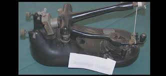
 Object 25: Cambridge rocker microtome
The Cambridge Rocker was one of the earliest automated microtomes. History. Before the invention of microtomes thin sections were cut for microscopy using a
 NATURE
A NEW ROCKING MICROTOME. SERIAL section-cutting has sprung into such The Rocking Microtome designed by the Cambridge. Scientific Instrument Company was ...
 Leica RM2255
Rotary Microtome. Instructions for Use. Leica RM2255 V 2.3 English 06/2018 completing a full handwheel rotation or to work in the rocking mode ("Rock").
 Leica RM2235 Rotary Microtome manual
The Leica RM2235 rotary microtome is equipped with a low-maintenance- slack-free micrometer drive
 YD-315 Rotary Microtome Operation Manual
It is used for slicing different level of thickness tissue specimen section for pathological diagnosis.. YD-315 rotary microtome is latest products which be
 HistoCore AUTOCUT R Rotary Microtome Instructions for Use
https://www.leicabiosystems.com/sites/default/files/media_product-download/2022-12/HistoCore_AUTOCUT_R_IFU_1v6I_en.pdf
 HistoCore NANOCUT R Rotary Microtome Instructions for Use V1.6
• Fully-Motorized Rotary Microtome with low-maintenance and backlash-free precision micrometer • Rocking mode function on the control panel for rapid trimming ...
 Rotary Microtome HM 355S
Operator Safety. • Safe ergonomic
 dical laboratory Techniques DepartmentMedical laboratory
Excluding ultra microtome there are 5 basic types. named according to the mechanism. — Rotary microtome. — Rocking microtome. — Rotary rocking microtome. —
 Object 25: Cambridge rocker microtome
Object 25: Cambridge rocker microtome. What is it? A microtome is an instrument used to cut very thin slices (sections) of tissue about half.
 Leica RM2125 RTS - Rotary Microtome
Rotary Microtome. Instructions for Use. Leica RM2125 RTS. V2.1 English - 10/2012. Order No. 14 0457 80101 RevB. Always keep this manual with the instrument
 Rocking Microtome PDF
The lead screw mechanism incorporates quality precision engineering and the cutting stroke is controlled by a single control lever which minimizes the
 YD-315 Rotary Microtome Operation Manual
It is used for slicing different level of thickness tissue specimen section for pathological diagnosis.. YD-315 rotary microtome is latest products which be
 dical laboratory Techniques DepartmentMedical laboratory
Excluding ultra microtome there are 5 basic types. named according to the mechanism. — Rotary microtome. — Rocking microtome. — Rotary rocking microtome.
 Leica RM2235 Rotary Microtome manual
Rotary Microtome. Instructions for use. Leica RM2235. V1.5; Rev B English – 01/2011. Order No. 14 0500 80101. Always keep this manual with the instrument.
 Microtomy - HM 340E
Rotary Microtome HM 340E. Stability and Speedy Preparation. • Highly stable blade holder platform yields high-quality ribbons even for.
 Microtome Safety Guidelines.pdf
Placement of the Blade. • Pay special precaution that a microtome blade is extremely sharp and must be handled cautiously. • Always set the rotary handle of the
 Rotary Microtome Microm HM 355S - 387861
Thermo Fisher Scientific. Operation Manual HM335S. 3. Microm HM355S. Rotary Microtome. Operation Manual – English ex Ser. No 44478
 Leica RM2145 - Rotary Microtome
Leica RM 2145 – Rotary Microtome. 1. Important information. Leica Microsystems Nussloch GmbH. Heidelberger Str. 17-19. D-69226 Nussloch. Germany.

HISTOLOGY AND CYTOLOGY
MODULEMicrotome
Histology and Cytology
50Notes9
MICROTOME
9.1 INTRODUCTION
A microtome (from the Greek mikros, meaning "small", and temnein, meaning "to cut") is a tool used to cut extremely thin slices of material, known as sections.OBJECTIVESAfter reading this lesson, you will be able to:
define Microtome describe the application of microtomes explain various microtomes describe microtome knife and its types.9.2 MICROTOME
Various types of microtomes are available. Most commonly used microtome for routine histopathology is rotary microtome.The most common applications of microtomes are:Traditional Histology Technique: Tissues are hardened by replacing water
with paraffin. The tissue is then cut in the microtome at thicknesses varying from 2 to 50 µm. From there the tissue can be mounted on a microscope slide, stained with appropriate aqueous dye(s) after prior removal of the paraffin, and examined using a light microscope. Cryosectioning Technique: Water-rich tissues are hardened by freezing and cut in the frozen state with a freezing microtome or microtome-cryostat; sections are stained and examined with a light microscope. This technique is much faster than traditional histology (15 minutes vs 16 hours) and is 51Microtome
HISTOLOGY AND CYTOLOGY
MODULE
Histology and Cytology
Notes used in conjunction with medical procedures to achieve a quick diagnosis. Cryosections can also be used in immuno histochemistry as freezing tissue stops degradation of tissue faster than using a fixative and does not alter or mask its chemical composition as much. Electron Microscopy Technique: After embedding tissues in epoxy resin, a microtome equipped with a glass or gem grade diamond knife is used to cut very thin sections (typically 60 to 100 nanometer). Sections are stained with an aqueous solution of an appropriate heavy metal salt and examined with a transmission electron microscope (TEM). This instrument is often called an ultramicrotome. The ultramicrotome is also used with its glass knife or an industrial grade diamond knife to cut survey sections prior to thin sectioning. These sections are of 0.5 to 1 µm thickness and are mounted on a glass slide and stained to locate areas of interest under a light microscope prior to thin sectioning for the TEM. Thin sectioning for the TEM is often done with a gem quality diamond knife. Botanical Microtomy Technique: Hard materials like wood, bone and leather require a sledge microtome. These microtomes have heavier blades and cannot cut as thin as a regular microtome. Rotary Mictrotome - It is most commonly used microtome. This device operates with a staged rotary action such that the actual cutting is part of the rotary motion. In a rotary microtome, the knife is typically fixed in a horizontal position. A rotary action of the hand wheel actuate the cutting movement. Here the advantage over the rocking type is that it is heavier and there by more stable. Hard tissues can be cut without vibration. Serial sections or ribbons of sections can easily be obtained. The block holder or block (depends upon the type of cassette) is mounted on the steel carriage that moves up and down and is advanced by a micrometer screw. Auto-cut microtome has built in motor drive with foot and hand control. With suitable accessories the machine can cut thin sections of paraffin wax blocks and0.5 to 2.0 micrometer thin resin sections.
Advantages
1. The machine is heavy, so it is stable and does not vibrate during cutting.
2. Serial sections can be obtained.
3. Cutting angle and knife angle can be adjusted.
4. It may also be used for cutting celloidin embedded sections with the help
of special holder to set the knife.HISTOLOGY AND CYTOLOGY
MODULEMicrotome
Histology and Cytology
52Notes
Fig. 9.1: Rotary MicrotomeFig. 9.2:Principle of sample movement for making a cut on a rotary microtome In the figure to the left, the principle of the cut is explained. Through the motion of the sample holder, the sample is cut by the knife position 1 to position 2), at which point the fresh section remains on the knife. At the highest point of the rotary motion, the sample holder is advanced by the same thickness as the section that is to be made, allowing for the next section to be made. The flywheel in microtomes can be operated by hand. This has the advantage that a clean cut can be made, as the relatively large mass of the flywheel prevents the sample from being stopped during the sample cut. The flywheel in newer models is often integrated inside the microtome casing. The typical cut thickness for a rotary microtome is between 1 and 60 µm. For hard materials, such as a sample embedded in a synthetic resin, this design of microtome can allow for good "Semi-thin" sections with a thickness of as low as 0.5 µm. Sledge Microtome is a device where the sample is placed into a fixed holder (shuttle), the sledge placed upon a linear bearing, a design that allows for the microtome to readily cut many coarse sections. Applications for this design of microtome are of the preparation of large samples, such as those embedded in paraffin for biological preparations. Typical cut thickness achievable on a sledge microtome is between is 10 and 60 micron.Fig. 9.3: MicrotomeFig. 9.4: A cryomicrotome
53Microtome
HISTOLOGY AND CYTOLOGY
MODULE
Histology and Cytology
NotesCryomicrotome
For the cutting of frozen samples, many rotary microtomes can be adapted to cut in a liquid nitrogen chamber, in a so-called cryomicrotome setup. The reduced temperature allows for the hardness of the sample to be increased, suchquotesdbs_dbs2.pdfusesText_3[PDF] roissy cdg terminal 4
[PDF] roissy en france nombre d'habitants
[PDF] roland barthes a lover's discourse analysis
[PDF] roland barthes a lover's discourse fragments
[PDF] roland barthes a lover's discourse fragments pdf
[PDF] roland barthes a lover's discourse pdf
[PDF] roland barthes quotes
[PDF] role of microorganisms in ecosystem slideshare
[PDF] roller coaster disneyland paris
[PDF] roman le langage secret des fleurs
[PDF] ronald dworkin
[PDF] roots of second order equation
[PDF] roskilde festival to do list
[PDF] rothschild india
