Dental canal anatomy

  • How many canals are in the teeth?

    The most common anatomy observed were: maxillary first premolars – 2 roots with 2 canals, maxillary second premolars – 1 root with 2 canals, mandibular first and second premolars – 1 root with 1 canal, maxillary first and second molars – 3 roots with 4 canals, and mandibular first and second molars – 2 roots with 3 .

  • What are the 5 basic root canal steps in order?

    The 5 Root Canal Procedure Steps

    1. Preparation.
    2. We will start by numbing the area with a local anesthetic.
    3. Accessing the Roots and Cleaning Them.
    4. Now, it's time for us to get into the root canals and pulp chamber.
    5. Shaping the Canals
    6. Filling the Canals
    7. Closing Off Access to the Canals

  • What are the different types of canals in dentistry?

    The three main types of root canal treatment based on materials used are traditional root canal treatment, non-surgical root canal treatment, and regenerative endodontics..

  • What is canal anatomy classification?

    Type I: a single main canal is present starting from the pulp chamber to the root apex.
    Type II: two separate canals leave the pulp chamber but join to form one canal to the apex.
    Type III: one canal leaves the pulp chamber and divides into two smaller canals which later merge again to exit through one canal..

  • What is the anatomy of the canal of the tooth?

    The root canal (or pulp canal) is one of the main parts of the anatomy of a tooth.
    It is located within the root which is the part of the tooth that extends below the gum.
    There is only one root canal in single-rooted teeth.
    In multirooted teeth there is one canal per root..

  • What is the process of a tooth canal?

    After space is cleaned and shaped, the endodontist fills the root canals with a biocompatible material, usually a rubber-like material called gutta-percha.
    The gutta-percha is placed with an adhesive cement to ensure complete sealing of the root canals.
    In most cases, a temporary filling is placed to close the opening..

  • What is tooth canal?

    A root canal treatment is a dental procedure to remove inflamed or infected pulp on the inside of the tooth which is then carefully cleaned and disinfected, then filled and sealed..

  • The three main types of root canal treatment based on materials used are traditional root canal treatment, non-surgical root canal treatment, and regenerative endodontics.
A root canal is the naturally occurring anatomic space within the root of a tooth. It consists of the pulp chamber (within the coronal part of the tooth), the main canal(s), and more intricate anatomical branches that may connect the root canals to each other or to the surface of the root.
The root canal contains the vessels and nerves that supply the teeth. Learn about it anatomy and function now at Kenhub!

What is a root canal?

The root canal is the portion of the pulp canal space within the root of the tooth limited by the pulp chamber and the foramen that follows the external outline of the root ( Fig

12 6 )

What is the significance of canal anatomy?

The significance of canal anatomy has been emphasized by studies demonstrating that variations in canal geometry before cleaning, shaping, and obturation procedures had a greater effect on the outcome than the techniques themselves

What is the structure and surrounding structures of a tooth?

Structure and surrounding structures of a tooth seen in cross section

The root canal (or pulp canal) is one of the main parts of the anatomy of a tooth

It is located within the root which is the part of the tooth that extends below the gum

There is only one root canal in single-rooted teeth

In multirooted teeth there is one canal per root

A root canal is the naturally occurring anatomic space within the root of a tooth. It consists of the pulp chamber (within the coronal part of the tooth), the main canal (s), and more intricate anatomical branches that may connect the root canals to each other or to the surface of the root.
Dental canal anatomy
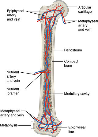
All bones possess larger or smaller foramina (openings) for the entrance of blood-vessels; these are known as the nutrient foramina, and are particularly large in the shafts of the larger long bones, where they lead into a nutrient canal, which extends into the medullary cavity.
The nutrient canal (foramen) is directed away from the growing end of bone.
The growing ends of bones in upper limb are upper end of humerus and lower ends of radius and ulna.
In lower limb, the lower end of femur and upper end of tibia are the growing ends.
The nutrient arteries along with nutrient veins pass through this canal.
A nutrient canal is found in long bones, in the mandible, and in dental alveoli.
In long bones the nutrient canal is found in the shaft.

Categories

Tooth anatomy canine
Oral anatomy canine
Dental anatomy chart canine
Oral cancer anatomy
Oral cancer anatomy affected
Dental root canal anatomy
Inferior dental canal anatomy
Canine dental anatomy terms
Dental anatomy app for pc
Dental in anatomy
Dental anatomy for the dental hygienist
Anatomy for dental students pdf free download
Anatomy for dental medicine pdf
Anatomy for dental students pdf
Anatomy for dental medicine
Anatomy for dental medicine 3rd edition pdf
Dentist maroc
Dental anatomy is a
Dental school anatomy requirements
Gross anatomy dental school