cell fixation protocol for sem
What is fixation in SEM?
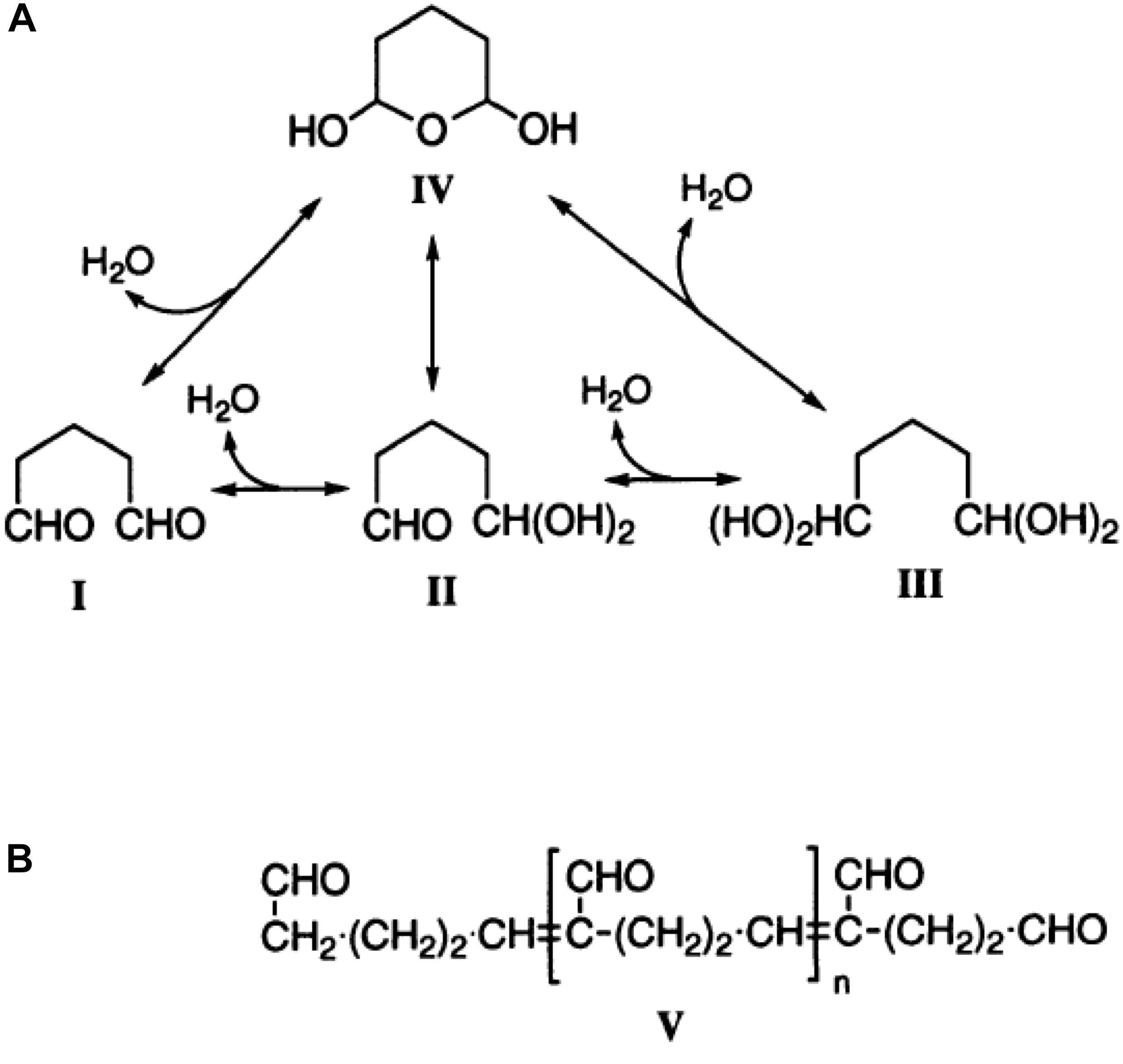
Fixatives containing both paraformaldehyde and glutaraldehyde provide a much better quality of fixation, than either aldehyde alone.
Formaldehyde penetrates tissues rapidly and mildly stabilizes proteins etc. which are then permanently fixed by the glutaraldehyde.How do you fix a SEM sample?
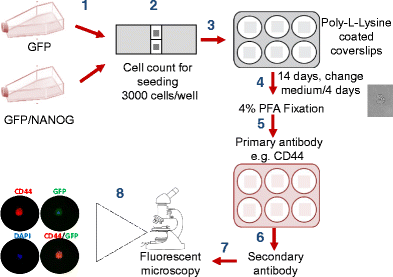
Fix specimens with an appropriate aldehyde fixative for a minimum of an hour.
Fixative routinely used by the Core and provided, 3% Glutaraldehyde in 0.
1) M Phosphate buffer.
For larger specimens can use 2% Paraformaldehyde/2% Glutaraldehyde in 0.
1) M Phosphate buffer.How do you fix cells for electron microscopy?
Fixation of tissues is the most crucial step in the preparation of tissue for observation in the transmission electron microscope.
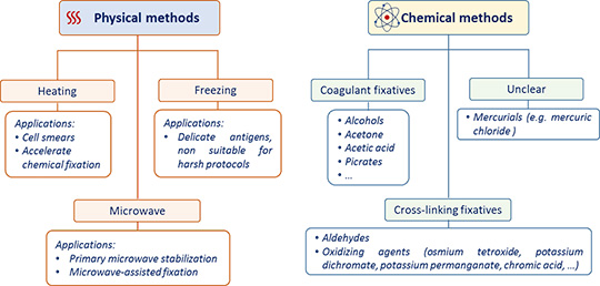
Fixation consists of two steps: cessation of normal life functions in the tissue (killing) and stabilization of the structure of the tissue (preservation).
- Fixation 1-2 hrs in 2% glutaraldehyde in 0.1M Sodium cacodylate buffer, pH 7.4.
- Rinse 3 X 10-15 min 30-45 min in 0.1M sodium cacodylate buffer, pH 7.4.
- Post-Fix 1-2 hrs in 1% Osmium tetroxide in water.
- Rinse 3 X 5 min in water.
- Dehydrate.
- Critical Point Dry 45-60 min or dry using HMDS.
|
Sample Preparations for Scanning Electron Microscopy – Life
solutions result in cell swelling and poor fixation. The most commonly used buffers for processing protocol and realized only at the SEM viewing stage. |
|
Scanning Electron Microscopy
need to be considered when selecting fixation protocols to reduce these vation of animal tissues and cells bacteria |
|
Scanning electron microscopy preparation and analysis of the cell
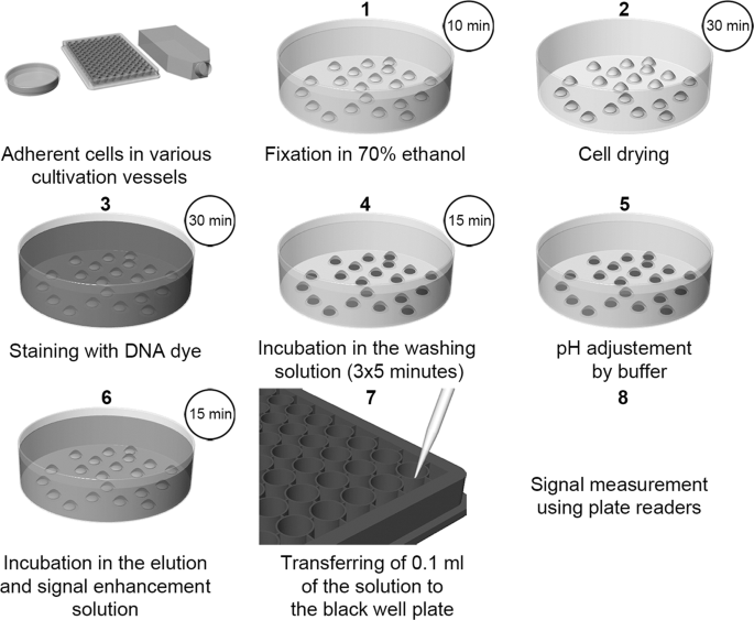
7 janv. 2020 Most SEM preparation protocols involve the crucial step of sample drying. ... for 30 min at 37 °C prior to cell pre-fixation step. |
|
A Modified Short Protocol for Preparation of Bryophytes for
fixation that may cause collapse of cell walls or other artifacts. The protocol reported here yielded acceptable specimens for SEM. |
|
TECHNICAL PROTOCOLS
4.2 Fixing cells in GrowDex-T with PFA [5]: . 5.2 Protocol for Scanning Electron Microscopy (SEM) of GrowDex and cells in GrowDex [7]: ........... 9. |
|
Methanol fixation for scanning electron microscopy of plants
ation as an alternative to standard protocols for SEM of plants. Methanol fixation stressed cell wall length dimensions of rye coleoptile seg-. |
|
Multi-photon Direct Laser Writing and 3D Imaging of Polymeric
7 juin 2017 different fixation protocols according to the desired imaging ... 2.2 Optical and SEM investigation of N2A cells morphology and seeding ... |
|
A Facile Method for Simultaneous Visualization of Wet Cells and
6 août 2020 Keywords: ionic liquid wet cell SEM |
|
New Insights Into Sperm Ultrastructure Through Enhanced Scanning
22 avr. 2021 which can be used in chemical fixation (SEM examination ... In addition this protocol makes cells dark enough to be. |
|
SPECIMEN PREPARATION PROTOCOL FOR TRANSMISSION
Specimen Preparation Protocol Fixing Cells for Electron Microscopy Scanning Electron Microscopy Protocol Using HMDS |
|
Sample Preparations for Scanning Electron Microscopy – Life
Fixation of samples is probably the most crucial step in SEM sample preparation protocols, b Protocol for Cultured Micro-organisms (Loose or Loosened Cells) |
|
Biological Specimen Preparation for Scanning Electron Microscope
Gentle liquid medium to bring the fixative to the cellular components • Help maintain normal pH levels while the cells are being fixed • Maintain osmotic |
|
A PROTOCOL FOR ENHANCED IMAGING AND - CORE
preparation technique of cervical cells for the FE-SEM/EDX study cells The proposed protocol was conducted by a McDowell-Trump fixative prepared in 0 1 M |
|
Scanning electron microscopy preparation and analysis of the cell
7 jan 2020 · Most SEM preparation protocols involve the crucial step of sample drying for 30 min at 37 °C prior to cell pre-fixation step Unless otherwise |
|
Preparation for SEM - Center for Microscopy and Image Analysis
Critical point drying Fixation Dehydration Coating SEM Critical point drying Fixation Freeze-fractured Vero cell: NO sublimation Cryo processing for SEM |
|
Osmium Tetroxide Staining for Cells - Electron Microscopy Sciences
Osmium Tetroxide is a good fixative and excellent stain for lipids in Visualized cellular structures depend on the fixation protocols; in Glutaraldehyde fixation |


