carti anatomie umana pdf
|
Human Anatomy
Associate Professor MDPhD Department of Anatomy and Embryology English Section Medicine First Year Victor Babeș University of Medicine and Pharmacy Timisoara |
|
Anatomia trunchiului si membrelor
3 Autori: Prof univ dr med SORIN-LUCIAN BOLINTINEANU Universitatea de Medicină și Farmacie „Victor Babeș” din Timișoara Conf univ dr med MONICA ADRIANA VAIDA Universitatea de |
Alina Maria Șișu
Associate Professor, MD,PhD, Department of Anatomy and Embryology, English Section, Medicine, First Year Victor Babeș University of Medicine and Pharmacy Timisoara old.umft.ro
Sorin Lucian Bolintineanu
Full Professor, MD,PhD, Head of Department of Anatomy and Embryology, Victor Babeș University of Medicine and Pharmacy Timisoara old.umft.ro
The layers of the eyeball
From external to internal there are three layers: I. A conjunctive layer, consisting of the sclera posterior and the cornea anterior; II. A vascular pigmented layer, formed of the choroid, ciliary body, and iris; III. A nervous layer, the retina. old.umft.ro
The sclera
The sclera is very dense and hard. It maintains the form of the eyeball. It is thicker posteriorly than anteriorly. Its external surface is white, opaque. Its anterior part is covered by the conjunctive membrane. Its internal surface is brown. It contains grooves, in which the ciliary nerves and vessels exist. Posteriorly it is pierced by the op
The cornea
The cornea is the transparent part of the external layer. It forms the anterior 1/6th of the surface of the eyeball. It is convex anteriorly, being dense and thick. The cornea is fromed of four layers: corneal epithelium, continuous with that of the conjunctiva; substantia propria posterior elastic lamina (lamina elastica posterior; De
The choroid (Chorioidea)
The choroid is a thin, vascularized membrane. It has got a dark brown colour. It forms the posterior 5/6th eyeball. It is travelled by the optic nerve. It is thicker posterior than anterior. Its external surface is closely situated to the sclera. Its internal surface is attached to the pigmented layer of the retina. The choroid consists of sm
(Corpus ciliare)
The ciliary body is formed of the followings: orbiculus ciliaris, ciliary processes, ciliaris muscle. The ciliary processes (Processus ciliares) are formed by the choroid, the choroid proper and the lamina basalis. The process is called the folding. They are arranged in a circle. They are attached by their periphery to the ridges of the orbiculu
The iris
The iris is a circular, contractile disk, suspended in the aqueous humor. It is located in between cornea and lens. It is pierced in the middle by a round aperture, the pupil. Its lateral part is continuous with the ciliary body. The iris separates the space between the lens and the cornea into an anterior and a posterior subspaces. They are call
3. The retina (Tunica interna)
The retina is a nervous membrane. Its role is of capturing the images of objects. Its external surface is in relations with the choroid. Its internal surface is in relation with the hyaloid membrane of the vitreous body. It is continuous posteriorly with the optic nerve. It decreases in thickness from back to front, until the ciliary bod
Retina proper
The elements of the retina proper are disposed in the layers as follows: Stratum opticum. Ganglionic layer. Internal plexiform layer. Internal nuclear granular layer. External plexiform layer. External nuclear granular layer. Layer of rods and cones. The stratum opticum represents the layer of nerve fibers. It consists of fibers of the optic nerve
4. The refracting media
The refracting media: Aqueous humor Vitreous body Crystalline lens. old.umft.ro
The aqueous humor (Humor aqueus)
The anterior and posterior chambers of the eyeball are filled with aqueous humor. It has got of water. old.umft.ro
The vitreous body (Corpus vitreum)
The vitreous body forms 4/5th of the eyeball. It is transparent and is composed of an albuminous fluid wrapped into the hyaloid membrane. In its center there is a canal, the hyaloid canal, containing lymph. The hyaloid membrane envelops the vitreous body. The portion anterior to the ora serrata has got radial fibers, forming the zonula ciliar
The crystalline lens (Lens crystallina)
The crystalline lens is situated posterior to the iris and anterior of the vitreous body. The capsule of the lens (Capsula lentis) is transparent. It is elastic and is situated in the hyaloid fossa in the anterior part of the vitreous body. It comes in relation anteriorly with the free border of the iris. It forms the posterior chamber of the eye
5. The accessory organs (Organa oculi accessoria)
The accessory organs of the eyeball are: the ocular muscles, the fasciæ, the eyebrows, the eyelids, the conjunctiva, the lacrimal apparatus. old.umft.ro
The muscles of the eyeball (Musculi oculi)
The ocular muscles are the: Levator palpebræ superioris Rectus superior Rectus inferior Rectus medialis. Rectus lateralis. Obliquus superior. Obliquus inferior The Levator palpebræ superioris muscle: It has origin on the inferior surface of the small wing of the sphenoid, superior and anterior to the optic foramen. At its origin ends anteriorly in
(Organon Auditus) * A. M. Șișu
The ear or organ of hearing is divided into three parts: the external ear, the middle ear or tympanic cavity, the internal ear or labyrinth. old.umft.ro
The external ear
The external ear has got a part named the auricula or pinna, and the external acoustic meatus. Pinna collects the vibrations of the air. The external acoustic meatus transmits the vibrations to the tympanic cavity. old.umft.ro
auricula or pinna
It has an ovoid form, its larger end superiorly. Its lateral surface is irregular and concave, presenting eminences and depressions. The elevation part of the auricula is named the helix. It presents a small tubercle, the auricular tubercle of Darwin. Parallel with the helix there is the antihelix. In between there is a triangular depression,
The external acoustic meatus (Meatus acusticus externus)
It extends from the concha until the tympanic membrane. It is directed internal, anterior, and superior (Pars externa). It passes internal and posterior (Pars media), and internal, anterior and inferior (Pars interna). The tympanic membrane closes the internal end of the meatus and has an oblique direction. The external acoustic meatus is forme
(Cavum tympani)
The middle ear or tympanic cavity is an irregular space located into the temporal bone. It contains air, conveyed from the nasopharynx into the auditory tube. It contains a chain of movable bones. They convey the vibrations reached to the tympanic membrane to the internal ear. The tympanic cavity consists of two parts: tympanic cavity proper,
The auditory ossicles (Ossicula auditus)
The tympanic cavity contains the following ossicles: malleus, incus, stapes. The malleus is attached to the tympanic membrane. The incus is placed in between. Both are connected bones via joints. The malleus or hammer, consists of a head, neck, and three processes: the manubrium, the anterior and the lateral processes. The head articulates p
Joints of the auditory ossicles
The incudo-malleolar joint is a saddle-shaped diarthrosis. It is covered by an articular capsule. The incudo-stapedial joint is an enarthrosis. It is covered by an articular capsule. Ligaments of the ossicles They are: The anterior ligament of the malleus The superior ligament of the malleus The lateral ligament of the malleus The posterior
(Auris interna)
The internal ear is the essential structure of the organ of hearing. Here there are found the final branches of the auditory nerve. It is named the labyrinth, due to complexity of its shape. It consists of two parts: the osseous labyrinth: there are spaces (in petrous part of the temporal bone), the membranous labyrinth: there are communicating
The Utricle (Utriculus)
The utricle, the biggest, occupies the superior and posterior part of the vestibule. Through its anterior wall exit the ductus utriculosaccularis. It opens into the ductus endolymphaticus. old.umft.ro
The Saccule (Sacculus)
The saccule is the smaller out of the two. Its anterior part presents the macula acustica sacculi, which receives the saccular filaments of the acoustic nerve. From the posterior wall a canal, the ductus endolymphaticus is united by the ductus utriculosaccularis. It passes along the aquæductus vestibuli, finishing in saccus endolymphaticus. The
4. The acoustic nerve (N. acusticus) or nerve of hearing or vestibulo-cochlear nerve
They are found in almost every part of the skin. They are situated in small pits on the inferior surface of the corium, in the subcutaneous areolar tissue, surrounded by adipose tissue. They are large in regions where the amount of perspiration is great, as in axillæ. They are large in the groin. They are a lot on the palms and on the soles.
|
Anatomia omului
Prima carte de anatomie în limba franceză “Anatomie univer- Este autorul primului tratat românesc complet de anatomie umană: “Manual practic de disecţie” şi “ ... |
|
A367.pdf
celulele sangvine ovulul |
|
CURRICULUM Calificarea profesională: Domeniul de pregătire
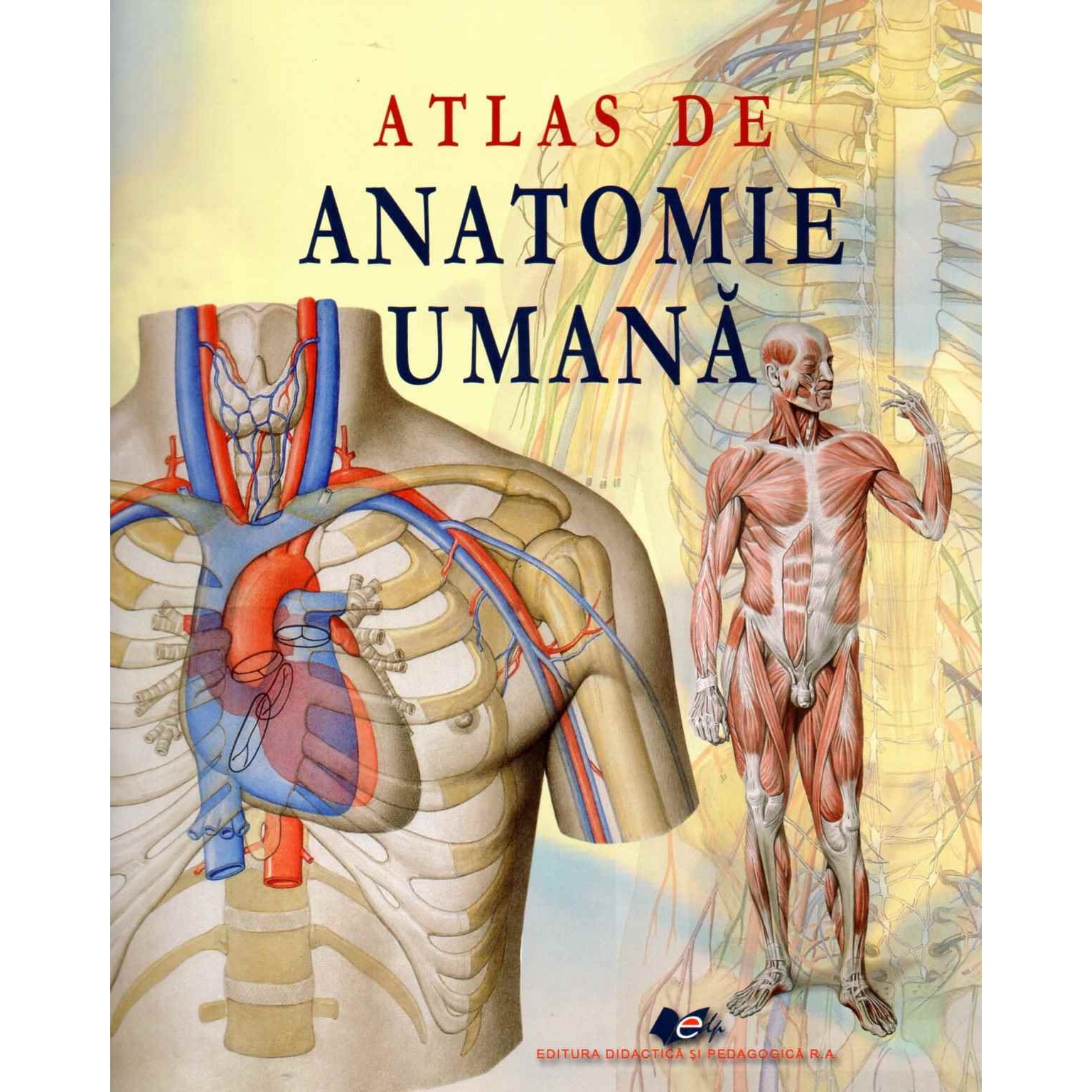
*** Atlas de anatomie umană Editura Didactică și Pedagogică |
|
Anatomia omului volumul I: Embriologie
embrionului uman ce duc la apariţia malformaţiilor congenitale. Conform desfăşurării în ordine cronologică |
|
ANDREAS VESALIUS ŞI ANATOMIA UMANĂ ÎN RENAŞTERE
demonstraţii publice de anatomie. După un an re- editează la Veneţia (1538) cartea de anatomie a ma- estrului său de la Paris Andernach: Institutiones. |
|
Manual de Anatomie şi Morfologie sportivă - Gheorghe Baciu
Convenţional în anatomie corpul uman este studiat în poziţie verticală cu perioadă) |
|
ANATOMIA OMULUI. Volumul II - Timișoara
nului dezvoltarea lui fiind caracteristică ortostatismului uman. Este un – Anatomie |
|
Examenul național de bacalaureat 2021 Proba E. d) Anatomie şi
Probă scrisă la anatomie şi fiziologie umană genetică şi ecologie umană. Model. Pagina 1 din 3. Examenul național de bacalaureat 2021. Proba E. d). Anatomie şi |
|
NOŢIUNI DE ANATOMIA ŞI FIZIOLOGIA OMULUI
Corpul uman este împărţit într-o serie de regiuni ce pot fi identificate la suprafaţa corpului. (fig.4). Regiunile mari ale corpului sunt: - capul. - gâtul. |
|
Examenul național de bacalaureat 2023 Proba E. d) Anatomie şi
28 iun. 2023 Probă scrisă la anatomie şi fiziologie umană genetică şi ecologie umană. Varianta 5. Barem de evaluare şi de notare. Pagina 1 din 2. Examenul ... |
|
Anatomia omului
Prima carte de anatomie în limba francez? “Anatomie univer- Tratatul s?u de anatomie uman? publicat în patru volume |
|
B A Z E L E A N A T O M I C E ALE MISCARII
Axe plane anatomice |
|
ANDREAS VESALIUS ?I ANATOMIA UMAN? ÎN RENA?TERE
editeaz? la Vene?ia (1538) cartea de anatomie a ma- estrului s?u de la Paris cu adev?rat ?tiin?ifice de anatomie uman? ?i totodat?. |
|
CURRICULUM Calificarea profesional?: Domeniul de preg?tire
Atlas de anatomie uman? Editura Didactic? ?i Pedagogic? |
|
Anatomia omului volumul I: Embriologie
Embriologia uman? este o ramur? a anatomiei ce studiaz? evolu?ia ontogenetic? a fiec?rui organism uman începând cu momentul fecund?rii. |
|
Programa-Biologie-bac-2021.pdf
Feb 6 2021 ANATOMIE ?I FIZIOLOGIE UMAN? |
|
S N?TOAS? ?
O alt? tendin?? ce se poate constata dac? privim alimenta?ia uman? din Dup? descoperirile de anatomie |
|
GRIGORE T. POPA ?I VICTOR PAPILIAN ÎNTRE „JURIU DE
blicase la Cluj un „Tratat de Anatomie uman?“ (2). Aprinse trebuie s? fi fost discu?iile în ilicite ?i plagieri (într-o carte menit? s? educe pe. |
|
Preciz?ri legate de terminologia folosit? în manualul de Anatomie ?i
4 Anatomie ?i fiziologie uman?. CAPITOLUL 5. Pagina. Localizare în text. În loc de: Se va citi: 104. Figura 6.4. Metacarpianul distal. Falanga distal?. |
|
Anatomia omului - USMF
Prima carte de anatomie în limba franceză “Anatomie univer- selle du corp complet de anatomie umană: “Manual practic de disecţie” şi “Tratatul elementar de |
|
ANATOMIA OMULUI - Catedra de anatomie și anatomie clinică
Prima carte de anatomie în limba franceză „Anatomie universelle du corp humain ” În anul 1897 a editat „Cursuri de anatomie practică”, iar în 1924 – „Manual de românești de anatomie umană, de embriologie, de antropologie și al unui |
|
Carte: Biologie – Anatomie si fiziologie umana, genetica si ecologie
Genetică umană Este ramura geneticii care studiază ereditatea şi variabilitatea populaţiei umane Genomul uman Genomul uman cuprinde integral informaţia |
|
ANATOMIE GENERALĂ
ANATOMIE GENERALĂ Alcătuirea corpului uman Cezar Th Niculescu, Radu Cârmaciu, (2008), Biologie - Manual pentru clasa a- XI-a, editura Corint |
|
Carte Biologie Anatomie Si Fiziologie Umana Genetica - str-tnorg
Acces PDF Carte Biologie Anatomie Si Fiziologie Umana Genetica Carte Biologie Anatomie Si Fiziologie Umana Genetica Right here, we have countless book |
|
PDF ANATOMIA OMULUI VOL 1 VICTOR PAPILIAN
Termenul „Anatomie” vine de la grecescul anatemnein, care înseamnă a tăia, a diseca Anatomia omului este ºtiinţa care studiază forma ºi structura organismului |
|
AnatomieNote de curspdf
ramură a anatomiei numită antropologia fizică sau anatomia generală Artiştii plasticieni au unei formaŃiuni anatomice, iar în anatomia dezvoltării are sensul de frontal; de fiinŃa umană, această durată medie a vieŃii a scăzut considerabil |


![Interiorul Corpului Uman Nr3 - [PDF Document] Interiorul Corpului Uman Nr3 - [PDF Document]](https://cdn4.libris.ro/img/pozeprod/59/1002/B8/833704.jpg)


