immunofluorescence protocol tissue
|
IMMUNOFLUORESCENCE
Miguel Angel Maestro and Mark Kalisz 2015. A-‐ Paraffin Blocks. 1 -‐ Dissect the tissue and place in PBS on ice (record weight number |
|
IHC-PARAFFIN PROTOCOL (IHC-P)
Since antibodies are highly specific the antibody will bind only to the protein of interest in the tissue section. The antibody-antigen interaction is then |
|
Abcam
1.5. Rinse briefly in phosphate-buffered saline (PBS). 2. Frozen (cryostat) sections. 2.1. Snap frozen fresh tissue in liquid |
|
Joyner Lab 2007 Protocol for immunofluorescent staining of mouse
Tissue: cryosections adhered to slides from blocks embedded in OCT using the 2- methylbutane (isobutene) method: see cryoprotection and processing of. |
|
Tissue preparation and cryopreservation with sucrose -- for frozen
16 Dec 2016 (See lacZ – β-galactosidase staining protocol for an alternative fixative that allows longer times). For immunostaining some antibodies can ... |
|
A Unique Immunofluorescence Protocol to Detect Protein
14 Jun 2017 Staining was performed on 10 glioma tissue sections along with 5 of their cryo sections 5 sections each of hepatocellular |
|
Chai Lab
Note: Air dry xylene slides in the hood and circle the tissue sections with. Hydrophobic Barrier PAP Pen b) 100% Ethanol------ 2-3 min RT. |
|
Methanol Fixation Immunofluorescence Staining & Imaging for
This protocol outlines methanol fixation immunofluorescence staining |
|
Methanol Fixation Immunofluorescence Staining & Imaging for
This protocol outlines methanol fixation immunofluorescence staining |
|
RNAscope® Multiplex Fluorescent v2 Assay combined with
Draw 2–4 times around tissue using the. ImmEdge™ hydrophobic barrier pen. Let the barrier dry ~30 SEC. ICW Pretreatment and Immunofluorescence. Prepare |
|
Abcam
1.5. Rinse briefly in phosphate-buffered saline (PBS). 2. Frozen (cryostat) sections. 2.1. Snap frozen fresh tissue in liquid |
|
Joyner Lab 2007 Protocol for immunofluorescent staining of mouse
Protocol for immunofluorescent staining of mouse frozen sections. Tissue: cryosections adhered to slides from blocks embedded in OCT using the 2-. |
|
IMMUNOFLUORESCENCE
IMMUNOFLUORESCENCE. Miguel Angel Maestro and Mark Kalisz 2015. A-? Paraffin Blocks. 1 -? Dissect the tissue and place in PBS on ice (record weight number |
|
Ten Approaches That Improve Immunostaining: A Review of the
26?/01?/2022 Consider adding steps 1–3 of the tissue preparation to your immunofluorescence protocol. 2.2.5. Disadvantages. (a) The relatively high cost of ... |
|
MULTIPLEX IMMUNOFLUORESCENCE PROTOCOL
Pre-incubate primary antibody with BSA (0.5%) prior to application to the tissue. •. Dilute primary antibody in antibody diluent to a working concentration. •. |
|
A Unique Immunofluorescence Protocol to Detect Protein
14?/06?/2017 Staining was performed on 10 glioma tissue sections along with 5 of their cryo sections 5 sections each of hepatocellular |
|
IHC-PARAFFIN PROTOCOL (IHC-P)
Since antibodies are highly specific the antibody will bind only to the protein of interest in the tissue section. The antibody-antigen interaction is then |
|
Chai Lab
Note: Air dry xylene slides in the hood and circle the tissue sections with. Hydrophobic Barrier PAP Pen b) 100% Ethanol------ 2-3 min RT. |
|
Methanol Fixation Immunofluorescence Staining & Imaging for
Ensure that tissue sections have been placed onto the appropriate slide prior to starting this Demonstrated Protocol. Consult the Visium Spatial Protocols - |
|
Optimized immunofluorescence staining protocol for imaging
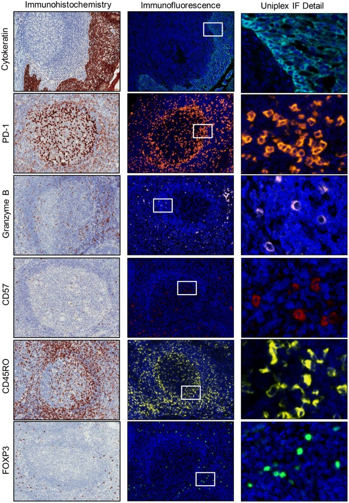
Location of immune cells that form the germinal center reaction within secondary lymphoid tissues can be characterized using confocal microscopy. |
|
Immunocytochemistry and immunofluorescence protocol - Abcam
Immunocytochemistry and immunofluorescence protocol Procedure for staining of cell cultures using immunofluorescence ICC and IF protocol Preparing the slide Coat coverslips with polyethylineimine or poly-L-lysine for 1 h at room temperature Rinse coverslips well with sterile H2O (three times 1 h each) |
|
IMMUNOFLUORESCENCE A GUIDE TO SUCCESSFUL IF
Immunofluorescence (IF) combines the use of antibodies with fluorescence imaging techniques to visualize target proteins and other biomolecules within fixed cell or tissue samples This process can reveal the localization relative expression and even activation states of target proteins |
|
Immunofluorescence for paraffin-embedded tissue sections
Immunofluorescence for paraffin-embedded tissue sections DAY 1 1) Remove paraffin and rehydrate the tissue sections a) Xylene -----2x 5min RT (** Perform this step in the hood**) Note: Air dry xylene slides in the hood and circle the tissue sections with Hydrophobic Barrier PAP Pen b) 100 Ethanol----- 2-3 min RT |
|
IMMUNOFLUORESCENCE A GUIDE TO SUCCESSFUL IF
Protocol for immunofluorescent staining of mouse frozen sections Tissue: cryosections adhered to slides from blocks embedded in OCT using the 2-methylbutane (isobutene) method: see cryoprotection and processing of embryonic tissue protocol This protocol is also suitable for 40µm free floating |
|
Tissue Cyclic Immunofluorescence (t-CyCIF) - protocolsio
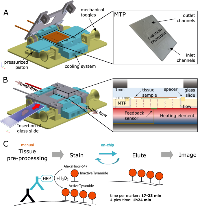
We describe a tissue-based cyclic immunofluorescence (t-CyCIF) method for highly multiplexed immunofluorescence imaging of specimens mounted on glass slides t-CyCIF generates up to 60-plex images using an iterative process (a cycle) in which conventional low-plex fluorescence images are repeatedly collected from the same sample and then |
|
Searches related to immunofluorescence protocol tissue filetype:pdf
Protocol for immunofluorescence staining of adhesion cells Protocol for immunofluorescence staining of adhesion cells This is provided as a general protocol Optimization of concentration or incubation condition of the primary antibody and the secondary antibody for your own specimen is necessary |
What is the immunofluorescence protocol?
- Immunofluorescence is a powerful tool for elucidating the complex signaling events that underlie biological processes and disease. This guide highlights critical steps in the immunofluorescence protocol and demonstrates how protocol changes can affect the final outcome of your experiment.
What is immunofluorescence staining?
- Immunofluorescence (IF) staining is a method of choice in studying the subcellular localization of proteins in fixed biological samples ( Zaglia et al., 2016; Niedenberger and Geyer, 2018; Smith and Gabriel, 2018 ).
What is immunofluorescence microscopy?
- Immunofluorescence microscopy is a powerful technique that is widely used by researchers to assess both the localization and endogenous expression levels of their favorite proteins. The application of this approach to C. elegans, however, requires special methods to overcome the diffusion barrier of a dense, collagen-based outer cuticle.
What is double immunofluorescence?
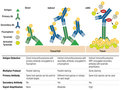
- Double immunofluorescence – simultaneous protocol In order to be able to examine the co-distribution of two (or more) different antigens in the same sample, a double immunofluorescence procedure can be carried out. Primary antibodies raised in different species can be used either in parallel (in a mixture) or in a sequential way.
|
IMMUNOFLUORESCENCE - Cell Signaling Technology
Immunofluorescence (IF) combines the use of antibodies with fluorescence imaging techniques to visualize target proteins and other biomolecules within fixed cell or tissue samples This process can reveal the localization, relative expression, and even activation states of target proteins |
|
Double immunofluorescence – simultaneous protocol - Abcam
1 5 Rinse briefly in phosphate-buffered saline (PBS) 2 Frozen (cryostat) sections 2 1 Snap frozen fresh tissue in liquid |
|
Joyner Lab 2007 Protocol for immunofluorescent staining of mouse
Tissue: cryosections adhered to slides from blocks embedded in OCT using sections cut on a vibratome (see protocol for free floating immunohistochemistry) |
|
Step-by-step protocol for whole mount immunofluorescence of
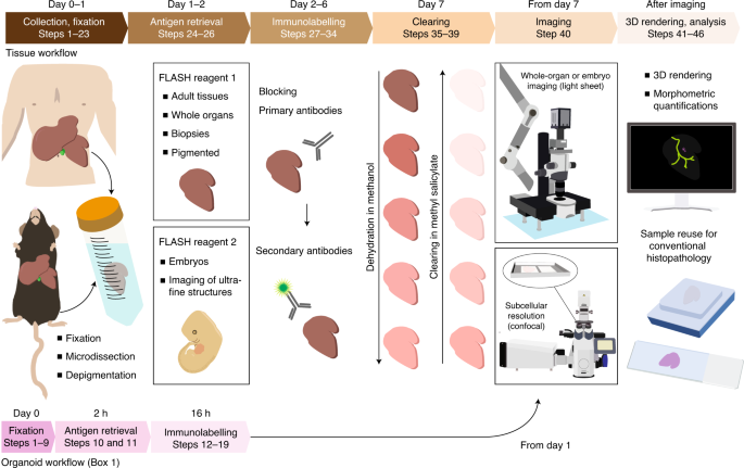
Dissect the tissue in ice-cold PBS Remove as much membranes as possible 2 Fix in fresh 4 paraformaldehyde (PFA) in PBS at 4oC for 2-3h Note: Old PFA |
|
IMMUNOFLUORESCENCE
IMMUNOFLUORESCENCE Miguel Angel Maestro and Mark Kalisz 2015 A-‐ Paraffin Blocks 1 -‐ Dissect the tissue and place in PBS on ice (record weight, |
|
MULTIPLEX IMMUNOFLUORESCENCE PROTOCOL
Pre-incubate primary antibody with BSA (0 5 ) prior to application to the tissue • Dilute primary antibody in antibody diluent to a working concentration |
|
Protocol 3: Immunofluorescence on Frozen Sections
23 mar 2015 · Remove tissue from sucrose and drain excess solution Place the tissue sample in the OCT compound, keeping track of tissue orientation Adjust |
|
Immunofluorescence Staining Protocol - St Michaels Hospital
Follow procedure for pretreatment as required 2 Pretreatments of Tissue Sections Page 2 Antigenic determinants masked by formalin-fixation |
|
IHC/ICC Protocol Guide - R&D Systems
Optimization of our staining protocols for tissue sections typi cally begin with an overnight incubation with the primary antibody at 4 °C For staining cells, a 1 hour |


