[PDF] principe d'échographie
[PDF] cryptography engineering design principles and pra
[PDF] cryptographie pdf
[PDF] applied cryptography
[PDF] decors chretiens de sainte sophie
[PDF] basilique sainte-sophie vikidia
[PDF] frise chronologique de sainte sophie
[PDF] chapelle du palais d'aix
[PDF] fonction d'une basilique
[PDF] plan de la basilique sainte sophie
[PDF] sainte sophie plan
[PDF] conseiller d'animation sportive salaire
[PDF] fiches ressources eps lycée professionnel
[PDF] conseiller technique sportif salaire
[PDF] programme eps lycée professionnel 2016
 167
167
[PDF] cryptography engineering design principles and pra
[PDF] cryptographie pdf
[PDF] applied cryptography
[PDF] decors chretiens de sainte sophie
[PDF] basilique sainte-sophie vikidia
[PDF] frise chronologique de sainte sophie
[PDF] chapelle du palais d'aix
[PDF] fonction d'une basilique
[PDF] plan de la basilique sainte sophie
[PDF] sainte sophie plan
[PDF] conseiller d'animation sportive salaire
[PDF] fiches ressources eps lycée professionnel
[PDF] conseiller technique sportif salaire
[PDF] programme eps lycée professionnel 2016
 167
167INTRODUCTION
Doppler echocardiography is a noninvasive tech-
nique that provides unique hemodynamic informa- tion otherwise not available without invasive moni- toring.The accuracy of the results depends,however, on meticulous technique and an understanding ofDoppler principles and flow dynamics.This docu-
ment provides recommendations based on the sci- entific literature and a consensus from a body of experts to guide the recording and measurement ofDoppler data.The document is not a comprehensive
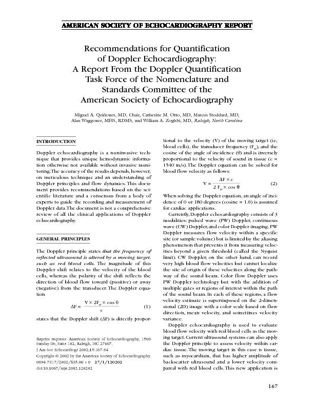
review of all the clinical applications of Doppler echocardiography.GENERAL PRINCIPLES The Doppler principle states that the frequency of reflected ultrasound is altered by a moving target, such as red blood cells.The magnitude of this Doppler shift relates to the velocity of the blood cells, whereas the polarity of the shift reflects the direction of blood flow toward (positive) or away (negative) from the transducer.The Doppler equa- tionΔF = ?
V×2F
o c×cosθ ?(1) states that the Doppler shift (ΔF) is directly propor- tional to the velocity (V) of the moving target (ie, blood cells), the transducer frequency (Fo ), and the cosine of the angle of incidence (θ) and is inversely proportional to the velocity of sound in tissue (c =1540 m/s).The Doppler equation can be solved for
blood flow velocity as follows: V =2FΔ
o F coc sθ (2) When solving the Doppler equation,an angle of inci- dence of 0 or 180 degrees (cosine = 1.0) is assumed for cardiac applications.Currently,Doppler echocardiography consists of 3
modalities: pulsed wave (PW) Doppler, continuous wave (CW) Doppler,and color Doppler imaging.PWDoppler measures flow velocity within a specific
site (or sample volume) but is limited by the aliasing phenomenon that prevents it from measuring veloc- ities beyond a given threshold (called the Nyquist limit). CW Doppler, on the other hand, can record very high blood flow velocities but cannot localize the site of origin of these velocities along the path- way of the sound beam. Color flow Doppler usesPW Doppler technology but with the addition of
multiple gates or regions of interest within the path of the sound beam.In each of these regions,a flow velocity estimate is superimposed on the 2-dimen- sional (2D) image with a color scale based on flow direction, mean velocity, and sometimes velocity variance.Doppler echocardiography is used to evaluate
blood flow velocity with red blood cells as the mov- ing target.Current ultrasound systems can also apply the Doppler principle to assess velocity within car- diac tissue.The moving target in this case is tissue, such as myocardium, that has higher amplitude of backscatter ultrasound and a lower velocity com-pared with red blood cells.This new application isReprint requests: American Society of Echocardiography, 1500
 Study on PRF Design and Doppler Measurement in Spaceborne