[PDF] Module Six - facksuedusa


Previous PDF Next PDF


Case Report Gestational Gigantomastia Complicating Pregnancy

peau d orange, and back pain Prolactin levels were g/L (with a normal reference value for prolactin in pregnancy being g/L) e patient was treated with bromocriptine ( mg twice daily), scheduled for a repeat cesarean, and referred to surgery for bilateral mammoplasty Conclusion


Widespread Tender Hemorrhagic Patches on the Breast

Nov 15, 2012 · changes in the breast area, such as peau d’orange, swell-ing, and redness Patients may also have itching, nipple occurs during pregnancy or breastfeeding Symptoms


24 The breast Their presence increases the chances of it, but

overlying localized ‘peau d’orange’ and fungation of tumour through the skin Mastitis carcinomatosa (rare) is a highly malignant form of carcinoma seen during pregnancy It is more generalized, and more like inflammation, or Burkitt's lymphoma, than the hard, fixed mass of a typical carcinoma


Management of breast cancer during pregnancy

or without a peau d’orange appearance This presentation is caused by tumour embolisation of dermal lymphatics The ‘milk-rejection’sign has been described rarely in case reports and refers to the unexplained refusal of an infant to feed from a lactating breast that harbours an occult carcinoma The differential diagnosis of a breast lump in


Module Six - facksuedusa

pregnancy or with significant weight gain or loss Redness is associated with breast inflammation A pigskin-like or orange-peel (peau d’orange) appearance results from edema, which is seen in metastatic breast disease The edema is caused by blocked lymphatic drainage 3- Inspect superficial venous pattern Observe visibility and pattern of


Anatomy 18 - Breast - WordPresscom

as peau d’orange (resembling an orange peel) In this case, connective tissue of suspensory ligaments shrinks, pulling the skin (which is the weakest part of the breast) and giving it the pitted appearance


Hyperthyroidism: Diagnosis and Treatment

Mar 01, 2016 · the tibiae with the skin assuming a peau d’orange (orange peel) appearance 18 Thyroid acropachy, an uncommon sign, is the clubbing of fingers and toes with soft-tissue swelling of the hands


Histology of Normal Breast - Elsevier Health

ligaments causes orange peel appearance of skin (peau d'orange) • Carcinoma involving these ligaments results in skin retraction &/or dimpling € Nipple • Positioned slightly medial and inferior to center of breast • 10-15 major lactiferous duct orifices open on surface of nipple Arranged radially in nipple


INFLAMMATORY BREAST CANCER

± warmth pain/itch enlargement peau d’ orange No recent pregnancy/lactation (non-puerperal) No response to antibiotics International IBC consensus, Ann

[PDF] peau d'orange treatment

[PDF] peau de chagrin analyse

[PDF] peau de chagrin balzac analyse

[PDF] peau de chagrin expression

[PDF] peau de chagrin larousse

[PDF] peau de chagrin personnages

[PDF] peau de chagrin résumé

[PDF] peau de chagrin résumé court

[PDF] peau de chagrin synonyme

[PDF] peau de triton au microscope

[PDF] pebble beach concours d'elegance 2016

[PDF] peche du carnassier en ille et vilaine

[PDF] peche intensive exposé

[PDF] péché originel bible

[PDF] Pêché originel d'Adam et Eve
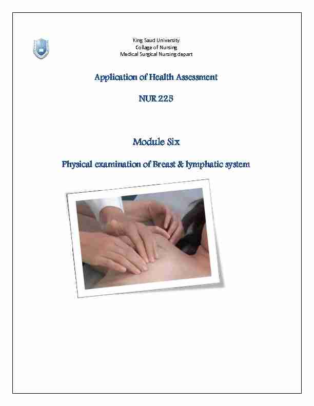
Module Six - facksuedusa

King Saud University

Collage of Nursing

Medical Surgical Nursing depart

Application of Health Assessment

NUR 225

Module Six

Physical examination of Breast & lymphatic system

I. Overview of the Anatomy

II. Purpose of Breast Examination

III. Obtaining Health History

IV. Physical Examination

V. Performance Checklist

I. Overview of anatomy:

- The breast also called mammary glands in women .lie on anterior chest wall.

- They are located vertically between the second or third and sixth ribs over the pectoralis muscle and

horizontally between the sternal border and the midaxillary line

- Each breast has centrally located nipple of pigmented erectile tissue ringed by an areola that darker

than tissue

OUTLINE

II. Purpose of breast examination:

9 To early detection and screening of breast cancer

9 To prevent breast cancer prevalence

9 To improve prognosis of breast cancer

9 To evaluate plan of care for breast cancer patient

III. Obtain health history:

Includes breast pain, nipple discharge, rash, lump, masses& other changes

Explore PAIN using COLDSPA:

Character: Describe pain (dull, aching, sharp)

Onset: When did this first begin?

Location: Point to the area where the pain occur. Ask whether it radiates to other areas. Duration: Ask how long does it last, recurrence and how often. Severity: Let patient describe pain using scale of 1-10 (10 being the most severe). Ask if it limits performance of activities of daily living. Pattern: What do you do when you have this pain? What medications do you take to relieve the pain? Associated Factors: Ask whether it occur at specific time during the menstrual cycle. Other associated symptoms (nipple discharge, changes in color of breast, swelling).

Chief complains

Age, day of menstrual cycle appears symptom, changes in underarm area axilla , tenderness , lump , or swelling , rash

Present history:

LMP( Last menstrual period ) , Delivery and pregnancy, breast feeding, history of breast disease, history of surgery or biopsy

Past history:

Breast cancer, breast disorder , other types of cancer Family history

Oral contraceptive pills Medication

High fat diet , caffeine Nutritional

Smoking, alcohol Habits

Perform breast self examination Self care behavior and psychological status

IV. Physical Examination

9 Equipment needed:

- Centimeter ruler - Small pillow - Gloves - Client handout for BSE - Slide for specimen

9 Prepare Patient:

- Explain the procedure in detail and answer any questions the client may have. - Provide privacy during the examination. - Work in a well lighted room. - Prepare the client for examination by having the client sit in an upright position. o Explain to the client that this is necessary to expose both breasts to compare for symmetry during the inspection. o One breast may be draped while the other breast is palpated. o The breasts are first inspected in the sitting position while the client is asked to hold arms in different positions. o The breasts are then palpated while the client assumes a supine position. - The final part of the examination involves teaching clients how to perform BSE and asking them to demonstrate what they have learned. Establish landmark and visualize the anatomy to describe clinical finding :

9 Method I: Divide the breast into four quadrants by

imaginary horizontal and vertical lines intersecting at the nipple. Describe findings according to: - Appropriate quadrant - The distance in centimeters from the nipple

9 Method II-Think of the breast as a clock with the nipple in

the centre. Describe findings according to: - Location according to time - The distance in centimeters from the nipple Remember: The upper outer quadrant of breast is the site of most breast tumors.

PHYSICAL EXAMINATION

FEMALE BREAST

ASSESSMENT PROCEDURE NORMAL FINDINGS ABNORMAL FINDINGS

INSPECTION

1- Inspect size and symmetry.

9 Have the client disrobe and sit with

arms hanging freely.

9 Explain what you are observing to

help ease client anxiety.

May vary in size.

Somewhat Around and

pendulous.

One breast may be

normally larger than the other

Older Adult

considerations:

The older client often has

more pendulous, less firm, and saggy breasts.

Recent increase in the size of one

breast may indicate inflammation or an abnormal growth.

2- Inspect color and texture.

9 Be sure to note client͛s oǀerall skin

tone when inspecting the breast skin.

9 Note any lesions.

Color varies depending on

the client͛s skin tone.

Texture is smooth, with no

edema.

Linear stretch marks may

be seen during and after pregnancy or with significant weight gain or loss.

Redness is associated with breast

inflammation.

A pigskin-like or orange-peel (peau

d͛orange) appearance results from edema, which is seen in metastatic breast disease.

The edema is caused by blocked

lymphatic drainage.

3- Inspect superficial venous pattern.

9 Observe visibility and pattern of

breast veins.

Veins radiate either

horizontally and toward the axilla (transverse) or vertically with a lateral flare (longitudinal).

Veins are more prominent

during pregnancy.

A prominent venous pattern may

occur as a result of increased circulation due to a malignancy. An asymmetric venous pattern may be due to malignancy.

4- Inspect the areolas.

9 Note the color, size, shape, and

texture of the areolas of both breasts.

Areolas vary from dark pink

to dark brown, depending on the client͛s skin tones.

They are round and may

Peau d͛orange skin, associated with

carcinoma, may be first seen in the areola. vary in size. Small

Montgomery tubercles are

present.

5- Inspect the nipples.

9 Note the size and direction of the

nipples of both breasts.

9 Also note any dryness, lesions,

bleeding, or discharge.

Nipples are nearly equal

bilaterally in size and are in the same location on each breast.

Nipples are usually

everted, but they may be inverted or flat.

No discharge should be

present.

Older Adult

Considerations

The older client may have

smaller, flatter nipples that are less erectile on stimulation.

A recently retracted nipple that was

previously everted suggestsquotesdbs_dbs2.pdfusesText_2