 ECG Basics - Boston College
Discuss a systematic approach to rhythm interpretation Review common cardiac arrhythmias Describe the process for interpretation of a 12 lead ECG
 The Basic 12 Lead Electrocardiogram
Teresa Camp-Rogers, MD Department of Emergency Medicine Virginia Commonwealth University Medical Center Richmond, Virginia The Basic 12 Lead Electrocardiogram
 The Electrocardiogram - UPSCALE
The Electrocardiogram The electrocardiogram (EKG or ECG) is a common diagnostic tool used to monitor heart activity In this experiment you will study the Physics of the EKG
 ECG INTERPRETATION:ECG INTERPRETATION
ECG INTERPRETATION:ECG INTERPRETATION: the basics Damrong Sukitpunyaroj MDDamrong Sukitpunyaroj, MD Perfect Heart Institue, Piyavate Hospital
 Électrocardiogramme (ECG ou EKG)
Electrocardiogram (ECG or EKG) rench Electrocardiogram (ECG or EKG) Électrocardiogramme (ECG ou EKG) An EKG is a test that records the electrical
 Recording a 12-lead electrocardiogram (ECG)
Place the first chest electrode (V1) in the fourth intercostal space, at the right sternal margin (Campbell et al , 2017) To identify this correctly, first locate the
 EKG - ECG CPT CODES - North Dakota
265 0-265 2 Thiamine and niacin deficiency states 270 0 Disturbances of amino-acid transport 272 0-272 9 Disorders of lipoid metabolism 274 82 Gouty tophi of other sites
 Électrocardiogramme
4e année médecine – Rotation 3 – 2015/2016 ISM Copy Module de Cardiologie Électrocardiogramme Généralités Enegiste l’actiité électi ue du cœu, c’est-à-die, la mesue et l’enegistement des potentiels
 Electrocardiogramme normal
Electrocardiogramme normal Année universitaire 2019-2020 1 Objectifs pédagogiques Objectif 1 : Connaitre la définition de l’électrocardiogramme
 Service Manual - Propaq CS Vital Signs Monitor
4 Safety summary Welch Allyn Propaq CS Vital Signs Monitor Keep away from rain Altitude limit Patient connections are Type B The device has met all essential requirements of European Medical Device Directive 93/42/
[PDF] phénomène périodique d'une paupière fermée
[PDF] business report example
[PDF] emploi du temps cm1 cm2 nouveaux programmes 2016
[PDF] report writing examples for students
[PDF] emploi du temps cm1 cm2 4 jours
[PDF] exemple d'emploi du temps cm1
[PDF] emploi du temps cm1 cm2 double niveau
[PDF] phénomène physique de la vie courante
[PDF] phenomene physique quotidien
[PDF] phénomènes physiques exemples
[PDF] effet leidenfrost
[PDF] rituels ce2
[PDF] rituels anglais collège
[PDF] introduction ? l'analyse des phénomènes sociaux pdf

The Electrocardiogram
The electrocardiogram (EKG or ECG) is a common diagnostic tool used to monitor heart activity. In this experiment you will study the Physics of the EKG.Prerequisites to this experiment are:
An understanding of the electric potential at about the level of OAC high school PhysicsDC Circuits I
Any experiment that uses an oscilloscope.
Background Information
The Electric Dipole
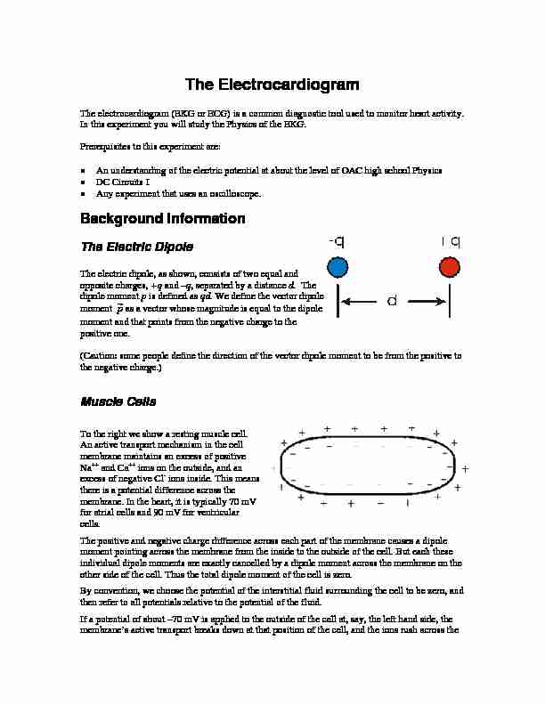
The electric dipole, as shown, consists of two equal and opposite charges, +q and -q, separated by a distance d. The dipole moment p is defined as qd. We define the vector dipole moment p as a vector whose magnitude is equal to the dipole moment and that points from the negative charge to the positive one. (Caution: some people define the direction of the vector dipole moment to be from the positive to the negative charge.)Muscle Cells
To the right we show a resting muscle cell.
An active transport mechanism in the cell
membrane maintains an excess of positive Na and Ca ions on the outside, and an excess of negative Cl ions inside. This means there is a potential difference across the membrane. In the heart, it is typically 70 mV for atrial cells and 90 mV for ventricular cells. The positive and negative charge difference across each part of the membrane causes a dipole moment pointing across the membrane from the inside to the outside of the cell. But each these individual dipole moments are exactly cancelled by a dipole moment across the membrane on the other side of the cell. Thus the total dipole moment of the cell is zero.By convention, we choose the potential of the interstitial fluid surrounding the cell to be zero, and
then refer to all potentials relative to the potential of the fluid.If a potential of about -70 mV is applied to the outside of the cell at, say, the left hand side, the
membrane's active transport breaks down at that position of the cell, and the ions rush across the 2 membrane to achieve equilibrium values. These equilibrium values turn out to be a slight excess of positive ions inside the cell, giving a potential inside the cell of about +10 mV; you may have learned about the Nernst equation in a Chemistry class that gives these equilibrium values. This potential difference at the left hand side of the cell causes the adjacent part of the cell membrane to similarly break down, which in turn causes its adjacent part to break down. Thus a wave of depolarisation sweeps down the cell from left to right with a velocity vas shown. Now the individual dipole moments across the cell membrane do not cancel each other out, and there is a net dipole moment of the cell pointing in the direction of the wave of depolarisation.When completely depolarised, the positive Ca
ion concentration inside the cell goes from about 7 105M to 6 105