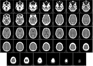
How can you tell the difference between a CT and MRI image?
CT scans use X-rays while MRI scans use strong magnets and radio waves.
A CT scan is generally good for larger areas, while an MRI scan produces a better overall image of the tissue under examination.
Both have risks but are relatively safe procedures..
How do MRI and CT scan technologies compare to each other?
Purpose: MRI scans are great for creating very detailed .
- D images of soft tissues, tendons, ligaments, your spinal cord and your brain.
CT scans are better suited for imaging injuries from trauma, staging cancer, and diagnosing conditions in blood vessels.
Is CT scan same as magnetic resonance?
MRI and CT scans use different technology to create diagnostic images of your body.
MRI uses radio waves, while CT uses X-rays.
CT scans are used to diagnose different conditions than MRI scans.
For example, if you had a suspected fracture, a CT scan is more suitable for finding the problem.Apr 5, 2023.
What is computed tomography and magnetic resonance imaging?
A CT scan is like a series of X-rays taken very quickly in a circle around you.
When combined and looked at together, they provide a detailed, three-dimensional image of your body.
MRIs use a large, powerful magnet and radio waves to create a similar picture..
What is the advantage of MRI imaging over computed tomography?
MRIs can create better pictures of organs and soft tissues, such as torn ligaments and herniated discs, compared to CT images..
What is the difference between a CT scan and an MRI scan?
MRI uses radio waves, while CT uses X-rays.
CT scans are used to diagnose different conditions than MRI scans.
For example, if you had a suspected fracture, a CT scan is more suitable for finding the problem.
They are also more suited to emergency situations as the results are quicker..
What is the difference between computed tomography and radiology?
A CT scan, or computed tomography scan, sends radiation through the body.
However, unlike a simple X-ray study, it offers a much higher level of detail, creating computerized, 360-degree views of the body's structures.
CT scans are fast and detailed.
They take longer than X-rays but are still fast (about one minute)..
What is the difference between CST and MRI?
What are MRIs and CT scans? A CT scan is like a series of X-rays taken very quickly in a circle around you.
When combined and looked at together, they provide a detailed, three-dimensional image of your body.
MRIs use a large, powerful magnet and radio waves to create a similar picture..
What is the difference between CT and MRA?
Magnetic resonance angiography vs.
Both of these are noninvasive imaging methods providers can use to see your blood vessels.
However, a CT (computed tomography) angiogram is faster than an MRA.
Also, they use different kinds of contrast materials.
A CT angiogram exposes you to radiation during the scan..
What is the difference between magnetic resonance imaging and computed tomography?
What are MRIs and CT scans? A CT scan is like a series of X-rays taken very quickly in a circle around you.
When combined and looked at together, they provide a detailed, three-dimensional image of your body.
MRIs use a large, powerful magnet and radio waves to create a similar picture..
Where are CT and MRI used?
MRI scans are generally considered as providing more accurate imagery and are therefore used for diagnosing conditions associated with your bones, organs or joints.
CT scans are often used to identify any bone fractures, tumours, or internal bleeding..
Where is magnetic resonance imaging used?
The brain, spinal cord and nerves, as well as muscles, ligaments, and tendons are seen much more clearly with MRI than with regular x-rays and CT; for this reason MRI is often used to image knee and shoulder injuries..
Why do we need magnetic resonance imaging?
Magnetic Resonance Imaging (MRI) is a non-invasive imaging technology that produces three dimensional detailed anatomical images.
It is often used for disease detection, diagnosis, and treatment monitoring..
Why is CT preferred to MRI?
Generally, CT scans are better at spatial resolution, while MRIs are better at contrast resolution.
That means CT scans are good at showing us where the edges of things are — where this structure ends and that other one begins..
- An MRI scan can produce more detailed images of tissues and organs than a CT scan.
An MRI scan may be used to diagnose issues with soft tissue, joints, organs, the brain and the heart.
MRI scans are more expensive than CT scans as the equipment is more costly and the process takes longer.Apr 5, 2023 - CAT scan: What's the difference? A CT scan and a CAT scan are the same thing.
CT stands for computerized tomography and CAT stands for computerized axial tomography.
The original name for this scan was an EMI scan, named after the company that created the technology. - CT scans take a fast series of X-ray pictures, which are put together to create images of the area that was scanned.
An MRI uses strong magnetic fields to take pictures of the inside of the body.
CT scans are usually the first choice for imaging.
MRIs are useful for certain diseases that a CT scan cannot detect.Jul 13, 2022 - Magnetic resonance imaging produces clearer images compared to a CT scan.
In instances when doctors need a view of soft tissues, an MRI is a better option than x-rays or CTs.
MRIs can create better pictures of organs and soft tissues, such as torn ligaments and herniated discs, compared to CT images.


