How is CT PNS test done?
CT PNS is an imaging technique that uses a rotating beam of X-rays to create cross-sectional images of the paranasal sinus (PNS).
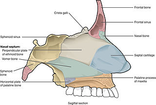
PNS are air-filled hollow spaces located between the bones of the skull and face.
CT PNS is helpful in detecting suspected disorders related to PNS..
What is a CT test for sinuses?
A computed tomography scan (CT or CAT) of your sinuses uses X-ray technology and advanced computer analysis to create detailed pictures of the sinus.
A CT of the sinus can help your physician to assess any injury, infection or other abnormalities..
What is CT scan of paranasal sinuses?
A computed tomography (CT) scan of the sinus is an imaging test that uses x-rays to make detailed pictures of the air-filled spaces inside the face (sinuses)..
What is the best scan for paranasal sinuses?
A CT scan gives good information about bony details and helps your doctor to decide whether the cancer is only in the sinuses or whether it has spread through the bone to surrounding structures..
What is the imaging of the paranasal sinuses?
Computed tomography (CT) and magnetic resonance imaging (MRI) are the primary imaging modalities for assessment of the paranasal sinuses.
Both these imaging techniques are far superior to plain radiographs in the evaluation of the sinonasal cavities..
What is the use of CT scan in PNS?
CT of the PNS is primarily used to: aid in the diagnosis of sinusitis.
Examine sinuses that have fluid in them or thickened sinus membranes.
By defining anatomy, you can plan for surgery..
What technique is used to examine the paranasal sinuses?
These are: plain X-ray, conventional tomography, computerised tomography (CT), and magnetic resonance (MR).
Plain X-ray is the initial examination, and is used as a screening procedure before employing one of the tomographic techniques..
What type of CT is used for sinuses?
A CBCT scan of the sinuses shows a patient's paranasal sinus cavities.
Paranasal sinuses are a group of four paired air-filled spaces that surround the nasal cavity.
With a CBCT scanner, our allergists are able to see the sinuses located under the eyes, above the eyes, between the eyes, and behind the eyes..
Why do I need a CT scan for nasal polyps?
CT scans can show the size of polyps deep in the sinuses and where they are.
These studies can also help rule out other reasons the nose is blocked.
Allergy tests.
Skin tests can show if allergies are causing ongoing inflammation..
Why do they do a CT scan of the sinuses?
CT rapidly creates detailed pictures of the sinuses.
The test may diagnose or detect: Abnormal nasal drainage.
An abnormal finding on an x-ray or nasal endoscopy..
- The scan specifically evaluates the paranasal sinus cavities, which are a group of four paired air-filled spaces that surround the nasal cavity.
Our x-rays expose the sinuses located under the eyes, above the eyes, between the eyes, and behind the eyes.
Unhealthy sinuses will show up as dark areas on the scan. - You will be asked to lie on a narrow table that slides into the center of the CT scanner.
You may lie on your back, or you may lie face-down with your chin raised.
Once you are inside the scanner, the machine's x-ray beam rotates around you.
You will not see the rotating x-ray beam.
