Computed tomography electron microscopy

  • How is electron microscopy done?

    Low voltage secondary electrons are emitted from the specimen surface and are attracted to the detector.
    The detector relays signals to an electronic console, and the image appears on a computer screen.
    Sometimes x-rays are detected and used to display the atomic elements within specimens..

  • How to do scanning electron microscopy?

    This is done by using a device called a "sputter coater." The sputter coater uses an electric field and argon gas.
    The sample is placed in a small chamber that is at a vacuum.
    Argon gas and an electric field cause an electron to be removed from the argon, making the atoms positively charged..

  • What does electron microscopy test for?

    Electron microscopy (EM) is a technique for obtaining high resolution images of biological and non-biological specimens.
    It is used in biomedical research to investigate the detailed structure of tissues, cells, organelles and macromolecular complexes..

  • What is electron microscope tomography?

    Electron tomography in biology is a technique to reconstruct the three-dimensional (.

    1. D) structure of macromolecular, subcellular, or cellular objects by transmission electron microscopy (TEM)

  • What is electron microscopy and electron tomography?

    Electron tomography (ET) is a tomography technique for obtaining detailed .

    1. D structures of sub-cellular, macro-molecular, or materials specimens.
    2. Electron tomography is an extension of traditional transmission electron microscopy and uses a transmission electron microscope to collect the data.

  • What is SEM imaging used for?

    Scanning electron microscopy (SEM) is a powerful technique for the analysis of a wide range of materials at high resolutions.
    SEM imaging relies on the detection of electrons which have been scattered from the surface and the bulk of a sample material after exposure to an electron beam..

  • What is the best reason for using a scanning electron microscope?

    The scanning electron microscope has many advantages over traditional microscopes.
    The SEM has a large depth of field, which allows more of a specimen to be in focus at one time.
    The SEM also has much higher resolution, so closely spaced specimens can be magnified at much higher levels..

  • What is the purpose of electron microscopy?

    Electron microscopy (EM) is a technique for obtaining high resolution images of biological and non-biological specimens.
    It is used in biomedical research to investigate the detailed structure of tissues, cells, organelles and macromolecular complexes..

  • What is the purpose of electron tomography?

    Electron tomography allows the study of the .

    1. D organization of thin individual cell organelles and bacterial cells at nanometer resolutions without slicing them.
    2. This technique is also able to reconstruct the .
    3. D structure of non-regular viruses

  • What is the TEM microscope used for?

    Transmission electron microscopes (TEM) are microscopes that use a particle beam of electrons to visualize specimens and generate a highly-magnified image.
    TEMs can magnify objects up to 2 million times.
    In order to get a better idea of just how small that is, think of how small a cell is..

  • What's the difference between TEM and SEM?

    The main difference between SEM and TEM is that SEM creates an image by detecting reflected or knocked-off electrons, while TEM uses transmitted electrons (electrons that are passing through the sample) to create an image..

  • Imaging methods in TEM use the information contained in the electron waves exiting from the sample to form an image.
    The projector lenses allow for the correct positioning of this electron wave distribution onto the viewing system.
  • The basic principle of the TEM is that a photographic image is recorded from the electron flux after it has passed through a thin sample of the specimen under study.
    The operating principle of TEM and image formation is illustrated schematically in Figure 8.10.
  • The TEM has the added advantage of greater resolution.
    This increased resolution allows us to study ultrastructure of organelles, viruses and macromolecules.
    Specially prepared materials samples may also be viewed in the TEM.
  • The transmission electron microscope is used to view thin specimens (tissue sections, molecules, etc) through which electrons can pass generating a projection image.
    The TEM is analogous in many ways to the conventional (compound) light microscope.
Electron tomography (ET) is a tomography technique for obtaining detailed 3D structures of sub-cellular, macro-molecular, or materials specimens.BF-TEM and ADF-STEM Atomic Electron Tomography
Electron tomography in biology is a technique to reconstruct the three-dimensional (3D) structure of macromolecular, subcellular, or cellular objects by transmission electron microscopy (TEM).
In a CT scan, the imaging equipment is moved around a patient to generate different images of a slice of the brain. In electron tomography, the specimen is tilted within the electron microscope to produce TEM images from many different perspectives to reconstruct a three-dimensional image.
Scanning electron microscopy (SEM) and micro-computed tomography (micro-CT) are high-resolution imaging modalities that enable visualization and quantification 
Computed tomography electron microscopy
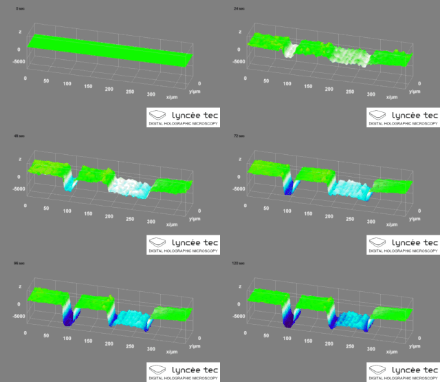
Digital holographic microscopy (DHM) is digital holography applied to microscopy.
Digital holographic microscopy distinguishes itself from other microscopy methods by not recording the projected image of the object.
Instead, the light wave front information originating from the object is digitally recorded as a hologram, from which a computer calculates the object image by using a numerical reconstruction algorithm.
The image forming lens in traditional microscopy is thus replaced by a computer algorithm.
Other closely related microscopy methods to digital holographic microscopy are interferometric microscopy, optical coherence tomography and diffraction phase microscopy.
Common to all methods is the use of a reference wave front to obtain amplitude (intensity) and phase information.
The information is recorded on a digital image sensor or by a photodetector from which an image of the object is created (reconstructed) by a computer.
In traditional microscopy, which do not use a reference wave front, only intensity information is recorded and essential information about the object is lost.
High-resolution transmission electron microscopy is an imaging mode of specialized transmission

High-resolution transmission electron microscopy is an imaging mode of specialized transmission

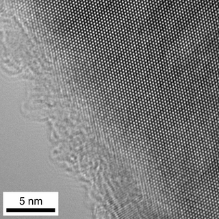
High-resolution transmission electron microscopy is an imaging mode of specialized transmission electron microscopes that allows for direct imaging of the atomic structure of samples.
It is a powerful tool to study properties of materials on the atomic scale, such as semiconductors, metals, nanoparticles and sp2-bonded carbon.
While this term is often also used to refer to high resolution scanning transmission electron microscopy, mostly in high angle annular dark field mode, this article describes mainly the imaging of an object by recording the two-dimensional spatial wave amplitude distribution in the image plane, similar to a classic light microscope.
For disambiguation, the technique is also often referred to as phase contrast transmission electron microscopy, although this term is less appropriate.
At present, the highest point resolution realised in high resolution transmission electron microscopy is around 0.5 ångströms (0.050 nm).
At these small scales, individual atoms of a crystal and defects can be resolved.
For 3-dimensional crystals, it is necessary to combine several views, taken from different angles, into a 3D map.
This technique is called electron tomography.

Categories

Computed tomography for mice
Computed tomography for bone mineral density
Micro-computed tomography (micro-ct) in medicine and engineering
Micro computed tomography in dentistry
Mic computed tomography
Micro-computed tomography (micro-ct)
Micro-computed tomography as a platform for exploring drosophila development
Nikon computed tomography
Computed tomography picture
Computed tomography pitch
Computed tomography pipe
Computed tomography pia
Computed tomography pins
Computerized tomography pictures
Computed tomography of pituitary
Electron beam computed tomography picture
Axial computed tomography of pilon fractures
Pipeline computed tomography
Computerized tomography risks
Computerized tomography ring