Cytology granuloma

  • How do you identify a granuloma?

    Your doctor or specialist will take a medical history and examine you if they suspect you have granulomas.
    They may ask for tests such as a blood test, x-rays or CT scans, genetic tests or a needle biopsy.
    With skin granulomas, your doctor may only need to do a physical examination to confirm a diagnosis..

  • What are the 5 types of granuloma?

    Six types of granulomatous skin lesions are identified according to cellular constituents and associated changes: 1) tuberculoid, 2) sarcoidal, 3) necrobiotic, 4) suppurative 5) foreign body and 6) histoid type granuloma (3,4)..

  • What is a granuloma histology?

    A granuloma is a focal aggregate of immune cells that forms in response to a persistent inflammatory stimulus.
    It characteristically demonstrates the compact organization of mature macrophages, which may or may not be associated with other inflammatory cell types..

  • What is a granuloma?

    A granuloma is a tiny cluster of white blood cells and other tissue.
    It can be found in the lungs, skin or other parts of the body.
    They form as a reaction to infections, inflammation, irritants or foreign objects.
    Granulomas aren't cancerous..

  • What is the histologic appearance of granuloma?

    Histologic subtypes.
    Granulomatous inflammation is commonly characterized by the formation of distinct granulomas composed of aggregates of epithelioid histiocytes, with a peripheral cuff of lymphocytes and plasma cells, and occasionally a necrotic center (Fig. 1)..

  • What is the process of granuloma?

    Granuloma formation begins with an inflammatory trigger, such as an infectious pathogen or a foreign body.
    Macrophages are recruited to the site of inflammation and activated as part of the innate immune response..

  • What type of cell is a granuloma?

    Granulomas are a tight collection of macrophages, often surrounded by helper T-cells.
    Most granulomas fall into one of two categories: caseating, with a necrotic center, or non-caseating, without any necrosis..

  • The following are important examples of conditions associated with granuloma formation.

    Tuberculosis. Sarcoidosis. Crohn Disease. Granulomatosis with Polyangiitis.
  • Conclusion: A significant number of cases of FNAC diagnosed granulomatous lymphadenitis have an identifiable underlying cause.
    Patients with reactive cytological changes, who clinically appear benign, can avoid unnecessary surgery.
  • Granulomas in cytological specimens from sarcoidosis patients are quite small [median (IQR) largest diameter: 0.478 (0.318–0.701) mm] and appear as well-defined, tiny nodules somehow 'popping out' of the slide, and they are easily appreciated even at low power ( fig.Dec 29, 2012
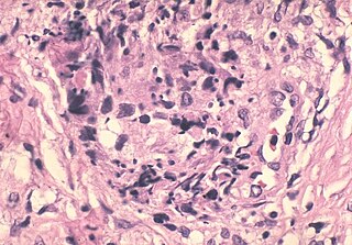
Fine needle aspiration cytology (FNAC) is very important investigation in the diagnosis of granulomatous inflammation. If it is supplemented with special stains like ZN, GMS, and PAS, it may help to differentiate between many infectious causes of granulomatous inflammation.
Abstract. Aims: Granulomatous mastitis (GM) is an uncommon breast lesion that mimics carcinoma. The fine needle aspiration cytological (FNAC) features of GM 

General

Definition of granuloma

,

How do I know if I have a granuloma?

Collection of cells that have abundant bubbly cytoplasm - most useful feature.
Multinucleated giant cells - these are easy to identify if you've seen some before.
Individual/singular multinucleated giant cells are not diagnostic of a granuloma.. but should raise one's suspicion of one being present.

,

Microscopic

Features that assist one in finding granulomas

,

Specific Sites

Lung granulomas

,

What is a granuloma?

Last Update:

  1. September 19
  2. 2022

A granuloma is a focal aggregate of immune cells that forms in response to a persistent inflammatory stimulus.
It characteristically demonstrates the compact organization of mature macrophages, which may or may not be associated with other inflammatory cell types.
,

What is chronic granulomatous disease?

There is a specific disease called chronic granulomatous disease; it is dealt with in the chronic granulomatous disease article.
Many definitions exist.
The term is used rather loosely by clinicans.
Radiologists occasionally call small lung nodules "granulomas".

,

What type of granuloma is a multinucleated giant cell?

Multinucleated giant cells are often present ( StatPearls:

  1. Granuloma [Accessed 13 June 2022] ) May present as a characteristic triad of florid follicular hyperplasia
  2. interfollicular monocytoid B cells and cortical / subcortical
  3. ill defined aggregates of epithelioid cells ( Toxoplasma lymphadenitis)

Medical condition

Cytology granuloma
Cytology granuloma

Lump of extravasated sperm found in some vasectomized men

A sperm granuloma is a lump of leaked sperm that appears along the vasa deferentia or epididymides in vasectomized individuals.
While the majority of sperm granulomas are present along the vas deferens, the rest of them form at the epididymis.
Sperm granulomas range in size, from one millimeter to one centimeter.
They consist of a central mass of degenerating sperm surrounded by tissue containing blood vessels and immune system cells.
Sperm granulomas may also have a yellow, white, or cream colored center when cut open.
While some sperm granulomas can be painful, most of them are painless and asymptomatic.
Sperm granulomas can appear as a result of surgery, trauma, or an infection.
They can appear as early as four days after surgery and fully formed ones can appear as late as 208 days later.

Categories

Cytology genetics evolution and plant breeding
Cytology genetics
Cytology grading system
Cytology genetics and evolution pdf
Cytology genetics and cytogenetics
Cytology guide
Cytology hindi
Cytology histology
Cytology hpv
Cytology hindi meaning
Cytology hpv testing
Cytology histiocytoma
Cytology history
Cytology histopathology
Cytology human anatomy definition
Cytology how to pronounce
Cytology histology and microscopic anatomy
Cytology how long for results
Cytology histology correlation
Cytology hospital