What is Multiplanar reconstruction (MPR)?
Multiplanar reconstruction (MPR) images of inflated and fixed lung in high-resolution (HR) mode (a–c) and non-HR mode (d–f). CT images were reconstructed with adaptive statistical iterative reconstruction (ASIR) (0%) (a,d), ASIR (40%) (b,e) and ASIR (80%) (c,f).
Does Multiplanar reconstruction of CT images increase the incidence of artefacts?
However, there was an increased incidence of artefacts by ASIR when CT images were obtained in non-HR mode. Multiplanar reconstruction (MPR) of CT images plays an important role in the interpretation of the three-dimensional anatomical location or extent of disease, and is an essential technique in daily clinical practice.
Can multiplanar Reformation (MPR) images be automatically generated on-the-fly?
We propose a system that automatically generates multiplanar reformation (MPR) images on-the-fly, which is independent of computed tomography (CT) scanner type.
How does multislice CT improve the quality of reconstructed MPRS?
Multislice CT has further improved the quality of MPRs as it allows isotropic imaging in which the image quality of the reconstructed MPR is the same as the original axial image. The axial plane passes through the body from anterior to posterior and divides it into superior and inferior sections.
|
Scanner multibarrette techniques reconstructions
Justifier l'utilisation des reconstruction 2D et 3D à partir d'acquisitions TDM. • Connaitre les impératifs d'acquisition. • Présenter les reconstructions |
|
Post-traitement en tomodensitométrie
Service de Radiologie A multiplanaires reformation multiplanaires. • Reconstruction à partir des coupes fines ? des données brutes. |
|
La pelvimétrie par scanner : état des connaissances actuelles
reconstruction multiplanaire. On travaille alors les coupes en reconstruction multiplanaire pour la mesure des diamètres habituels du bassin (cf. plus haut). |
|
Influence des données cliniques sur lanatomie trachéo-bronchique
25 avr. 2017 examen scannographique après reconstruction multiplanaire. ... Tous les patients ont bénéficié d'un scanner thoracique en fin d'expiration ... |
|
Case series
30 juin 2016 Tous ces patients ont été explorés par la radiographie standard un complément scanner avec reconstruction multiplanaires avant et après ... |
|
Comment je fais un phlébo-scanner cave supérieur ?
17 nov. 2011 Scanner thoracique : coupe axiale (a) et reconstruction multiplanaire coronale (b) montrant un envahissement tumoral de la veine cave supérieure ... |
|
Case report
24 mars 2015 Le scanner cervico-dorsal sans injection de produit de contraste avec reconstruction multiplanaire montrait une discrète majoration de la. |
|
Titre du mémoire
2.3.7 Etude CT-scan pour les fractures costales . 4.4.1 Réformation ou reconstruction multiplanaire sur GE et OsiriX .................. 58. |
|
CONE BEAM EN IMPLANTOLOGIE ORALE (Première partie
.la tomodensitométrie ou scanner dont les indications reposent aujourd'hui LOGICIELS DE RECONSTRUCTION MULTIPLANAIRES: Equipant tous. |
What is Multiplanar reconstruction CT scan?
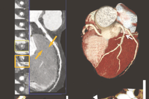
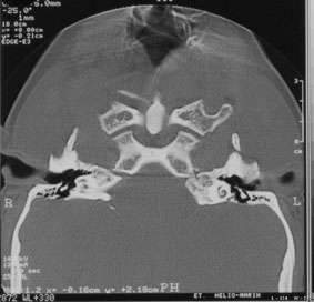
Multiplanar reconstruction (MPR) is the process of converting data from one anatomical planes (usually transverse) to other planes. It can be used for thin slices as well as projections. Multiplanar reconstruction is possible as present CT scanners provide almost isotropic resolution.
What is Multiplanar reconstruction (MPR)?
© 2013 Medixant 3.4Multiplanar reconstructions (MPR) The MPR tool provided within the RadiAnt DICOM Viewer can be used to reconstruct images in orthogonal planes (coronal, sagittal, axial or oblique, depending on what the base image plane is). This can help to create a visualization of the anatomy which was not possible using base images alone.
What is multi planar reconstruction in image processing?
Multiplanar Reconstruction. Multiplanar reconstruction or reformatting is a post-processing technique to create new images from a stack of images in planes other than that of the original stack. The use of thin slices increases the spatial resolution in the scan axis direction, allowing a high spatial resolution in all planes.
What are multiplanar reformations?
" Multiplanar reformations (MPR's) are two-dimensional reformatted images that are reconstructed secondarily in arbitrary planes from the stack of axial image data. " (Prokop and Galanski, 2003) 2003 Prokop, M., and M. Galanski.
| Quantifying the variability between multiple multiplanar |
| Evaluation of Multiplanar Reconstruction in CT Recognition |
| Basic Stages of PET Introduction to PET Image Reconstruction |
| Searches related to reconstruction multiplanaire scanner filetype:pdf |
Are multiplanar reconstructions useful for the diagnosis of lumbar disk disease?
- Axial computed tomographic (CT) images were compared with sagittal and coronal reformations and myelograms in 60 patients to evaluate the diagnostic usefulness of multiplanar reconstructions for the recognition of lumbar disk disease.
. The axial CT scans were most sensitive and specific.
What are the advantages of multi planar reconstruction?
- Such multi planar reconstructions have been helpful in the diagnosis of a variety of conditions including disk disease [8, 9].
. An additional advantage that has been claimed for this technique is that the lumbar CT examination can be entirely standardized.
What is iterative reconstruction in CT scanning?
- CT scanners used to rely on filtered back projection (FBP) for image reconstruction.
. Credited with fast processing times due to simplifying assumptions about the image chain, FBP had been the industry standard for almost 30 years.
. Today, iterative reconstruction (IR) algorithms/methods have emerged as the primary technique in reconstruction.
What is the best technique for image reconstruction?
- reconstruction (IR) algorithms/methods have emerged as the primary technique in reconstruction.
. The primary advantages of IR are its ability to incorporate attenuation corrections, reduce image noise, model the scanner itself, and even
|
Scanner multibarrette techniques reconstructions
Justifier l'utilisation des reconstruction 2D et 3D à partir d'acquisitions TDM • Connaitre les impératifs d'acquisition • Présenter les reconstructions principales |
|
Post-traitement en tomodensitométrie
Service de Radiologie A Hôpital Cochin - Paris multiplanaires, reformation multiplanaires • Reconstruction à partir des coupes fines ≠ des données brutes |
|
Imagerie Médicale 3D Visualisations, segmentations et
reconstruction utilisés en radiologie moderne, dont nous verrons rapidement particulière est connue sous le nom de reconstruction multi-planaire ou multi- |
|
Technique Scanner par JCP
Le scanner utilise des rayons X qui traversent le corps et sont plus ou moins reconnaître et de reconstruire les acquisitions perpendiculairement à l'axe de Les reconstructions multiplanaires ; les reconstructions se font dans tous les plans |
|
426 le scanner muItibarrette - Ce document est le fruit dun long
leurs soirées passées à nos côtés àla console du Scanner Nous tenons Reconstruction partielle (demi-tour) en acquisition séquentielle 2 2 1 2 Les reconstructions multiplanaire permettent une étude des coronaires dans les différents |
|
Apport des techniques de reconstruction en - ScienceDirect
Mots-clés : Imagerie thoracique Reconstruction Scanner SUMMARY Contribution of rie en coupes multiplanaires alors que celles de l'ère tomographique |
|
Imagerie en coupes le scanner volumique thoracique - ONCLE PAUL
dans l'axe Z de l'épaisseur de coupe (et il n'y a plus,sur les scanners modernes de possibilité par contre on choisit l'épaisseur de reconstruction par sommation de coupes fines ( si on ''épaissi'' les les reformations multiplanaires frontales |
|
Limagerie actuelle du thorax en tomodensitométrie - Edimark
Les dernières générations de scanners multi- coupes ou épaisseur de reconstruction d'environ 1 mm et une La technique du rendu volumique multiplanaire |
|
Evaluation de la prescription de la Tomodensitométrie aux urgences
15 oct 2011 · Service de radiologie des urgences 1- Evaluation des indications du scanner par les spécialistes 28 d) Reconstruction multiplanaire |
|
FACULTE DE MEDECINE DE TOURS - Université de Tours
3 avr 2015 · Exemple : Scanner thoracique, reconstruction multiplanaire, début d'acquisition en C7, car moitié du corps vertébral et pédicules de C6 non |
Can Computed Tomographic Gastrography and Multiplanar
reconstruction, For the first scan in the supine position 110-120 mL Visipaque 270 (iodixanol; GE) were given intravenously, and the tube current was 365 mA The abdomen was scanned from the diaphragm to the pubic symphysis 80 seconds after the start of the contrast injection at 3 mL/s for the portal venous phase in the first 10 patients
Scanner osseux fœtal 3D : technique, indications, limites
la reconstruction multiplanaire (figures 1 à 6) Les reconstructions multiplanaires 2D, segment par segment, permettent de confirmer les images 3D Pour cet examen, la patiente est généralement placée en décubitus dorsal ; le scanner peut être réalisé dès 24 SA, voire quelques semaines avant, toujours après discussion pluridisciplinaire
PROTOCOLE D’IMAGERIE MEDICALE
Une reconstruction multiplanaire dirigée sur les anomalies détectées est recommandée Pour la recherche de nodules pulmonaires, un post-traitement en reconstruction de type MIP (maximum intensity projection) d’une épaisseur d’environ 5 mm est impératif
FICHE UE2 - COURS 8 - IMAGERIE HYBRIDE
(reconstruction multiplanaire), MIP (projection d’intensité maximale) et VRT (technique de rendu volumique) Uniquement pour l’hybride (SPECT-TDM, TEP-TDM, TEP-IRM) on retrouve la mire de triangulation et le fVRT (fused volume rendering technique) MultiPlanar Representation (MPR) : Reconstruction multiplanaire
ZipFix pour sternum Une nouvelle
reconstruction multiplanaire avec une épaisseur de coupe de 22 mm 0X6 001 282_AA 02 11 11 15:28 Seite Cvr3 examen scanner et des techniques d'imagerie numérique
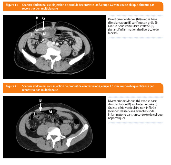
Perforation spontanée d’un diverticule de Meckel
scanner révèle une majoration des signes inflammatoires du diverticule associée à l’apparition d’un épaississement pariétal inflammatoire de contiguïté du côlon transverse (Figure 3) Figure 3 : Scanner abdominal avec injection de produit de contraste iodé, coupe 1 5mm, coupe frontale obtenue par reconstruction multiplanaire
LES TECHNIQUES DE RADIOLOGIE CONVENTIONNELLE LA RADIOGRAPHIE
quel que soit l¶axe de reconstruction, il procure moins d’artéfacts métalliques, notamment à l¶étage radiculaire et enfin il est au moins aussi précis que le scanner Scanner Cone beam Fig 2 11 Comparaison des deux explorations tridimensionnelles: scanner et cone beam Les mesures sont identiques
IMAGERIE CARDIOVASCULAIRE
Informatiques de reconstruction Cest un examcn très pefformant pour l'étude du poumon ct du médiastin, et des gros vaisseaux thoraciques (aortiques ct pulmonaires Les machines les plus récentes (64 barrettes ou plus), permettent d'examiner le cœur, ct les Coronaircs, a Princöoe
Le mésentère dans tous ses replis – L’épiploon dans toutes
des modes de reconstruction permettant de réduire les doses L’IRM est et devrait plus encore être utilisée en complément de l’échographie pour étudier les pathologies mésentériques, épiploïques et péritonéales Les atouts principaux de la technique sont son caractère non irradiant et la possibilité d’une approche multiplanaire
IMAGERIE EN IMPLANTOLOGIE ORALE - Dentalespacecom
préféré rester fidèles au scanner dans certaines indications, optimisant son utilisation en diminuant la dose émise, qui savère acceptable dans des conditions particulières (scanner 64 barrettes, tension, temps de pose et intensité minima) Le scanner, qui reste incontournable dans certaines indications et le cone beam, examen de première
|
Scanner multibarrette techniques reconstructions
[PDF] Scanner multibarrette techniques reconstructions sfrnet Data %reconstruction%DES pdf |
|
optimisation en pratique scanographique - ONCLE PAUL
[PDF] optimisation en pratique scanographique ONCLE PAULonclepaul RADIOPROTECTION FILEminimizer pdf |
|
Imagerie Médicale 3D Visualisations, segmentations et
[PDF] Imagerie Médicale D Visualisations, segmentations et bnazarian free MyUploads ImagerieMedicaleD pdf |
|
Scanner
[PDF] Scanner imre ucl ac be rpr RDGN scanner pdf |
|
L imagerie actuelle du thorax en tomodensitométrie - Edimark
[PDF] L 'imagerie actuelle du thorax en tomodensitométrie Edimark edimark Front frontpost getfiles pdf |
|
La pelvimétrie par scanner : état des connaissances - Edimark
[PDF] La pelvimétrie par scanner état des connaissances Edimark edimark Front frontpost getfiles pdf |
|
Intérêt de la reconstruction 3D préopératoire des vaisseaux - sfctcv
[PDF] Intérêt de la reconstruction D préopératoire des vaisseaux sfctcv sfctcv ftp journal jo pdf |
|
MC PROCEDURES SCANOGRAPHIQUES STANDARDISEES MC
[PDF] MC PROCEDURES SCANOGRAPHIQUES STANDARDISEES MC eassa cordo pagesperso orange SFROPRI scanMC pdf |
|
Comment je fais un phlébo-scanner cave supérieur - Club Thorax
[PDF] Comment je fais un phlébo scanner cave supérieur Club Thorax clubthorax PHLEBOSCANNER%CAVE%SUPERIEUR pdf |
|
01/12/2015 GUELORGET Alice CR : Kévin Boué Bases - AEM2
déc La radiographie et le scanner visualisent la valeur des numéros atomique des atomes constituants Reconstruction multiplanaire (MRP) |
- reconstruction mip irm
- fenetrage scanner
- reconstruction vrt
- vrt scanner
- reconstruction mip definition
- principe du scanner médical
- scanner monobarrette
- cours scanner radiologie
imagerie 3D en tomodensitométrie

imagerie 3D en tomodensitométrie

Le cœur en scanner thoracique : mon VADeMecuM - ScienceDirect

Imagerie Master UE Sciences Morphologiques PDF Téléchargement Gratuit

imagerie 3D en tomodensitométrie

John Libbey Eurotext - Hépato-Gastro \u0026 Oncologie Digestive

imagerie 3D en tomodensitométrie

John Libbey Eurotext - Hépato-Gastro \u0026 Oncologie Digestive

Le cœur en scanner thoracique : mon VADeMecuM - ScienceDirect

Postprocessing in Maxillofacial Multidetector Computed Tomography

Une baisse d'acuité visuelle récente -Reconstruction multiplanaire

Le cœur en scanner thoracique : mon VADeMecuM - ScienceDirect

Scanner cardiaque et coroscanner : quelles nouveautés en 2020

La Tomodensitométrie (scanner X)

Le cœur en scanner thoracique : mon VADeMecuM - ScienceDirect

La Tomodensitométrie (scanner X)

Comment je fais un scanner simple énergie pour embolie pulmonaire

Localisation de nodules pulmonaires en VATS grâce au Cone Beam

La Tomodensitométrie (scanner X)

imagerie 3D en tomodensitométrie

imagerie 3D en tomodensitométrie

Le cœur en scanner thoracique : mon VADeMecuM - ScienceDirect

Imagerie Master UE Sciences Morphologiques PDF Téléchargement Gratuit

imagerie 3D en tomodensitométrie

John Libbey Eurotext - Hépato-Gastro \u0026 Oncologie Digestive

imagerie 3D en tomodensitométrie

John Libbey Eurotext - Hépato-Gastro \u0026 Oncologie Digestive

Le cœur en scanner thoracique : mon VADeMecuM - ScienceDirect

Postprocessing in Maxillofacial Multidetector Computed Tomography

Une baisse d'acuité visuelle récente -Reconstruction multiplanaire

Le cœur en scanner thoracique : mon VADeMecuM - ScienceDirect

Scanner cardiaque et coroscanner : quelles nouveautés en 2020

La Tomodensitométrie (scanner X)

Le cœur en scanner thoracique : mon VADeMecuM - ScienceDirect

La Tomodensitométrie (scanner X)

Comment je fais un scanner simple énergie pour embolie pulmonaire

Localisation de nodules pulmonaires en VATS grâce au Cone Beam

La Tomodensitométrie (scanner X)

![imagerie 3D en tomodensitométrie imagerie 3D en tomodensitométrie]()
imagerie 3D en tomodensitométrie
![imagerie 3D en tomodensitométrie imagerie 3D en tomodensitométrie]()
imagerie 3D en tomodensitométrie
![Le cœur en scanner thoracique : mon VADeMecuM - ScienceDirect Le cœur en scanner thoracique : mon VADeMecuM - ScienceDirect]()
Le cœur en scanner thoracique : mon VADeMecuM - ScienceDirect
![Imagerie Master UE Sciences Morphologiques PDF Téléchargement Gratuit Imagerie Master UE Sciences Morphologiques PDF Téléchargement Gratuit]()
Imagerie Master UE Sciences Morphologiques PDF Téléchargement Gratuit
![imagerie 3D en tomodensitométrie imagerie 3D en tomodensitométrie]()
imagerie 3D en tomodensitométrie
![John Libbey Eurotext - Hépato-Gastro \u0026 Oncologie Digestive John Libbey Eurotext - Hépato-Gastro \u0026 Oncologie Digestive]()
John Libbey Eurotext - Hépato-Gastro \u0026 Oncologie Digestive
![Le cœur en scanner thoracique : mon VADeMecuM - ScienceDirect Le cœur en scanner thoracique : mon VADeMecuM - ScienceDirect]()
Le cœur en scanner thoracique : mon VADeMecuM - ScienceDirect
![Postprocessing in Maxillofacial Multidetector Computed Tomography Postprocessing in Maxillofacial Multidetector Computed Tomography]()
Postprocessing in Maxillofacial Multidetector Computed Tomography
![Une baisse d'acuité visuelle récente -Reconstruction multiplanaire Une baisse d'acuité visuelle récente -Reconstruction multiplanaire]()
Une baisse d'acuité visuelle récente -Reconstruction multiplanaire
![Le cœur en scanner thoracique : mon VADeMecuM - ScienceDirect Le cœur en scanner thoracique : mon VADeMecuM - ScienceDirect]()
Le cœur en scanner thoracique : mon VADeMecuM - ScienceDirect
![Scanner cardiaque et coroscanner : quelles nouveautés en 2020 Scanner cardiaque et coroscanner : quelles nouveautés en 2020]()
Scanner cardiaque et coroscanner : quelles nouveautés en 2020
![La Tomodensitométrie (scanner X) La Tomodensitométrie (scanner X)]()
La Tomodensitométrie (scanner X)
![Le cœur en scanner thoracique : mon VADeMecuM - ScienceDirect Le cœur en scanner thoracique : mon VADeMecuM - ScienceDirect]()
Le cœur en scanner thoracique : mon VADeMecuM - ScienceDirect
![La Tomodensitométrie (scanner X) La Tomodensitométrie (scanner X)]()
La Tomodensitométrie (scanner X)
![Comment je fais un scanner simple énergie pour embolie pulmonaire Comment je fais un scanner simple énergie pour embolie pulmonaire]()
Comment je fais un scanner simple énergie pour embolie pulmonaire
![Localisation de nodules pulmonaires en VATS grâce au Cone Beam Localisation de nodules pulmonaires en VATS grâce au Cone Beam]()
Localisation de nodules pulmonaires en VATS grâce au Cone Beam
![La Tomodensitométrie (scanner X) La Tomodensitométrie (scanner X)]()
La Tomodensitométrie (scanner X)
![Perforation spontanée d'un diverticule de Meckel </p>
</figure>
</div>
<br/>
<br/>
<script>
var imgs = document.querySelectorAll( Perforation spontanée d'un diverticule de Meckel </b></h3></figcaption>
<p>Source: Louvain Médical]()

imagerie 3D en tomodensitométrie

imagerie 3D en tomodensitométrie

Le cœur en scanner thoracique : mon VADeMecuM - ScienceDirect

Imagerie Master UE Sciences Morphologiques PDF Téléchargement Gratuit

imagerie 3D en tomodensitométrie

John Libbey Eurotext - Hépato-Gastro \u0026 Oncologie Digestive

imagerie 3D en tomodensitométrie

John Libbey Eurotext - Hépato-Gastro \u0026 Oncologie Digestive

Le cœur en scanner thoracique : mon VADeMecuM - ScienceDirect

Postprocessing in Maxillofacial Multidetector Computed Tomography

Une baisse d'acuité visuelle récente -Reconstruction multiplanaire

Le cœur en scanner thoracique : mon VADeMecuM - ScienceDirect

Scanner cardiaque et coroscanner : quelles nouveautés en 2020

La Tomodensitométrie (scanner X)

Le cœur en scanner thoracique : mon VADeMecuM - ScienceDirect

La Tomodensitométrie (scanner X)

Comment je fais un scanner simple énergie pour embolie pulmonaire

Localisation de nodules pulmonaires en VATS grâce au Cone Beam

La Tomodensitométrie (scanner X)

reconstruction vrt
Real-time tracking and navigationsystem for needle insertion
- reconstruction multiplanaire scanner
- filtre de reconstruction scanner
- reconstruction mip scanner
- cours scanner medical pdf
- reconstruction mpr
- reconstruction mip definition
- reconstruction mip irm
- cours scanner manip
reconstruction mip irm
Scanner multibarrette techniques reconstructions
- reconstruction mip scanner
- reconstruction 3d scanner
- reconstruction multiplanaire scanner
- filtre de reconstruction scanner
- reconstruction mip definition
- cours scanner medical pdf
- reconstruction vrt
- multiplanar reconstruction
imagerie médicale 3d
Imagerie Médicale 3D Visualisations, segmentations et
- reconstruction 3d scanner
- reconstruction mpr
- reconstruction mip scanner
- reconstruction mip definition
- reconstruction mip irm
- reconstruction vrt
- reconstruction multiplanaire scanner
- multiplanaire définition
démonstration fonction inverse dérivée
Fonctions dérivées - Académie en ligne
- fonction dérivée formule
- fonction dérivée tableau
- fonction dérivée exercice corrigé
- fonction dérivée 1ere s
- calcul de dérivée exercices corrigés
- fonction dérivée graphique
- fonction dérivée cours
- dérivée d'une fraction