 Yoga Anatomy
UNDERSTANDING THE ASANAS. 29. 3C. H APTER. Deciding which anatomical details of yoga poses to depict is quite a challenge. Unlike weight training and stretching
 Yoga Teacher Training Anatomy of Asanas in Hatha Yoga
Unlike the first anatomy manual that focused on the functions of specific muscle groups and then mentioned a few yoga postures that would stretch or strengthen
 Skeletal Muscle Anatomy
Introduction. As a yoga teacher it's important to have an understanding of how yoga asanas affect specific muscles
 Yoga as Experiential Learning in Undergraduate Anatomy Courses
yoga poses using only skeletal anatomy cues. Participants remained in each posture as the anatomy was reinforced. Next participants learned muscular
 Module 2 connective tissue and muscular system
How might you apply what you noticed to creating a feeling of grounding and support in standing asanas? Page 14. Instructor Guide. Module 8: The Anatomy of
 Zach Beach
another Yoga Anatomy Coloring Book). ○ Leslie Kaminoff Yoga Anatomy. ○ Ray Long
 Anatomy-of-Asana-I.compressed.pdf
start yoga. □ Commonly a provocative movement pattern – Flexion vs. Extension. □ Type of pain?
 A Critical Review Study on Importance of Anatomical Knowledge
Knowledge of anatomical principles can aid in understanding the beneficial effects of practicing Yoga Asana. Keywords: Ayurveda; Yoga Asana; Anatomical
 The-complete-guide-to-yin-yoga-philosophy-and-practice.pdf
Yoga and anatomy are intimately linked and Paul's investigations in these two Her yoga style blends both a Yin sequence of long-held poses to enhance the.
 Functional Anatomy of the Hamstring Muscle and Its Correla- tion
Although a few research studies have quantified the hamstring muscle activities in various yoga asanas evidence correlating it to functional anatomy is scarce.
 Yoga Anatomy
YOGA. ANATOMY. Leslie Kaminoff. Asana Analysis by. Amy Matthews. Illustrated by how the breath and spine come together in the practice of yoga postures.
 Yoga Teacher Training Anatomy of Asanas in Hatha Yoga
Unlike the first anatomy manual that focused on the functions of specific muscle groups and then mentioned a few yoga postures that would stretch or strengthen
 An Anatomical Illustrated Analysis of Yoga Postures Targeting the
Sep 30 2020 Key Words: Cadaveric dissection; Anatomical illustrations;. Back musculature; Yoga postures; Chronic lower back pain.
 Module 2 connective tissue and muscular system
Apply: In what other yoga poses do you think you could use this same technique to create make up the "anatomical stirrup" of the foot.
 Anatomy-of-Asana-I.compressed.pdf
start yoga. ? Commonly a provocative movement pattern – Flexion vs. Extension. ? Type of pain?
 yoga-anatomy-2nd-edition-pdfdrive.com_.pdf
Yoga anatomy / Leslie Kaminoff Amy Matthews ; Illustrated by Sharon by avoiding reductionist analysis of the poses and prescriptive listings of their ...
 Yoga as Experiential Learning in Undergraduate Anatomy Courses
Anatomy (YA) workshop instructors discussed muscle names locations
 anatomy of an asana
Extended Sides Angle Pose or 'Utthita. Parsvakonasana' as it is traditionally known is one of these asanas and will be taught in most yoga classes.
 RYA Brochure3_print
A 200-hours comprehensive Yoga teacher training certification course anatomy for each and every Yoga asana covered in this course.
 Instructor Guide
Explain the limitations of describing movement from anatomical position. Most yoga postures aren't single movements but instead they are a combination ...
Yoga Teacher Training
Anatomy of Asanas in Hatha Yoga
By: Nancy Wile
Yoga Education Institute
© Yoga Education Institute, 2014
All rights reserved. Any unauthorized use, sharing, reproduction or distribution of these materials by any means is strictly prohibited. 1Table of Contents
Introduction 2
Review of the Spine 3
Revi 6
Review of Biomechanics 9
Reflexes Related to Stretching 12
Imbalances 15
Anatomy of Standing 16
Anatomy of Specific Standing Postures 19
Anatomy of Sitting 29
Anatomy of Specific Seated Postures.. 32
Anatomy of Kneeling.. 37
Anatomy of Specific Kneeling Postures.. 39
Anatomy of Arm Balancing 42
Anatomy of Specific Arm Balancing Postures 43
Anatomy of Belly Lying / Lying Face Down 52
Anatomy of Specific Prone Postures 52
Anatomy of Back Lying / Lying Face Up 54
Anatomy of Specific Supine Postures 55
Anatomy of As 62
2Introduction
In this section of anatomy of hatha yoga, we will be going more in depth in our understanding of the foundations, biomechanics of movement, and muscles most affected in specific yoga postures. As we noted in the previous manual on anatomy, the spine mainly moves in four directions: 1) flexion (forward bending),2) extension (back bending), 3) lateral flexion (side bending), and 4) rotation.
In yoga, we have six main foundational (starting) points for any posture: 1) standing, 2) seated, 3) kneeling, 4) arm balancing, 5) prone (front lying), and 6) supine (back lying). In this manual, we will focus on the general anatomy for each starting point, and then examine postures from each of those foundational points that, grouping them by the final position of the spine in that posture (neutral, flexion, extension, lateral flexion, and rotation). We focus on the muscles most affected and how to properly move into postures based on their foundational starting point. We will examine the general anatomical consideration of each posture and how to use that knowledge to deepen the posture and avoid injury. The first sections will provide a review of the spine, biomechanics and major muscles groups that you learned about in the first anatomy of yoga manual (from the 200 hour program). Unlike the first anatomy manual that focused on the functions of specific muscle groups and then mentioned a few yoga postures that would stretch or strengthen each group, this manuals looks in detail at specific yoga asanas and the anatomy of movement related to each one. 3Review of the Spine
The Spine and Pelvic Girdle
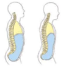
The spine has four distinct segments, consisting of the cervical, the thoracic, the lumbar, and the sacral. Each spinal segment contains a given number of vertebrae. The cervical spine has seven vertebrae, the thoracic (mid back) has twelve vertebrae, and the lumbar (low back) has five vertebrae. The vertebrae are separated by the intervertebral discs. These discs absorb shock, permit some compression, and allow movement. There are no discs in the sacrum or coccyx where the vertebrae are fused together. The cervical spine is curved in an extended position (cervical lordosis). The thoracic spine is curved in a flexed position (thoracic kyphosis). And the lumbar spine is curved in an extended position (lumbar lordosis). When there is too much rounding in the thoracic spine (like a hump back), it is call kyphosis.When there is too much arching in the
low back (lumbar region), it is called lordosis. When the spine is curved from right to left (rather than straight), it is called scoliosis. The sacrum is the foundation platform on which the spinal column is balanced. It is attached to the two hip bones at the sacroiliac joint. The rib cage consists of the twelve ribs that attach to the thoracic spine.The appendicular skeleton (including
shoulders, arms, pelvic girdle, legs) joins the axial skeleton (spinal column) at the shoulders and hips. The collar bone (clavicle) and the shoulder blade (scapula) form the shoulder. Each hip consists of three fused bones: the ilium, ischium, and pubis. This forms the pelvic girdle, which is shaped like a bowl. 4Neck and Spine
The portion of the spine contained within the neck is called the cervical spine. Unlike the rest of the spine, which is better protected from injury because it is enclosed by the torso, the cervical spine is more vulnerable to injury. This portion of the spine is enclosed in a small amount of muscles and ligaments, but is required to have extensive range of motion. People often experience neck strain due to repetitive or prolonged neck extension or flexion, often caused by poor posture while sitting or standing. It can also be caused by common habits, such as cradling a phone between the shoulder and ear, or sleeping in an awkward position. By gently strengthening and stretching the neck muscles, strain is reduced. Remember to not roll the head when stretching the neck. 5Deep Spinal Muscles (Neck/Back) Posterior View
Below is a diagram of the deep muscles of the spine. The group of long muscles along the spine are known as erector spinae. The erector spinae is made up of the iliocostalis, longissimus, and spinalis muscles. 6Review of Muscles
Muscle Forms
Muscles have different forms and fiber arrangements, depending on their function. Muscles in the limbs tend to be long. Because of this, they can contract more and are capable of producing greater movement. Muscles in the trunk tend to be broader and to form sheets that wrap around the body. Muscles that stabilize parts of the body tend to be short and squat, like those found in the hip. Muscles are also defined by the number of joints they cross; from their origin to their insertion. Monoarticular muscles cross only one joint, while polyarticular cross more than one joint (for example hamstrings).Types of Muscle Contractions
Muscles are composed of bundles of fibers held together by very thin membranes. Within these fibers are thousands of tiny filaments, which slide along each other when the muscle is stimulated by a nerve. This causes the muscle to shorten or contract. Muscles that produce a specific movement are called agonists, while the muscles that produce the opposite movement are called antagonists. When we think of a muscle contracting, we tend to think of the muscle shortening as it generates force. While this is one way that muscles contract, there are also other forms of muscle contraction.Concentric Contractions
In this form of contraction, a muscle shortens in length while contacting. An example is when the biceps brachii muscle in the forearm contracts to lift a book off a table and bring it in close to you to read, or when you perform a bicep curl with a free weight.Eccentric Contractions
When you slowly extend your elbow to put a book you were reading back on a table, you are lengthening the muscle (biceps brachii) while keeping some of its muscle fibers in a state of contraction. Whenever this happens, we call this movement eccentric lengthening; increasing muscle length against resistance or gravity.Isometric Contraction
In isometric contraction, muscles are active while held at a fixed length. The muscle is neither lengthened or shortened, but is held at a constant length. An example of isometric contraction would be carrying an object in front of you. The weight of the object would be pulling down, but your hands and arms would be opposing the motion with equal force going upwards. Since your arms are neither raising or lowering, your biceps will be isometrically contracting. 7Review of Major Muscles
The chart below is a review of the major muscles you learned about from your previous anatomy module you received during your 200 hour program. 8Psoas Muscle
The psoas major muscle contributes to hip flexion and it part of a group of muscles known as the hip flexors. 9Review of Biomechanics
Muscular Force in Yoga
When we align the long axis of the bones with the direction of gravity, we decrease the necessary muscular force to maintain a posture. This makes the posture feel more effortless. For example, when sitting in a cross leg position, gravity is aligned with the long axis of the spine. If we sit up tall, stacking the head over the spine, our muscles are less strained, than by rounding our backs and dropping our heads.Forward bends
Forward bends stretch and strengthen the back portion of the spine, pelvic girdle, shoulders and legs. They also strengthen the abdominal muscles, which contract as we bend forward, and gently compress abdominal organs, which stimulates their function.Proper Technique in Bending Forward
rounded, with the neck in line with the rest of the spine. So, as you bend forward you maintain t back and hinge from the hips, instead of rounding the back, when folding forward. If hamstrings are tight, it is best to bend the knees, so the back can remain flat. Rounding the back due to tight hamstrings can lead to a backwards rotation of the pelvis and a collapsing of the chest over the belly. This can result in intervertebral disc compression in the anterior (front) of the spine. Low back pain is often a result of poor mechanical relationship between the lumbar spine and the pelvis. Although many chronic back conditions can occur due to improper forward bending, forward bending done properly can also help us strengthen and stabilize our back and body. Obstacles to forward bends result from tightness in the hamstrings, spinal muscles and gluteals.Back Bends
Backward bending helps to stretch the front portion of the torso, shoulders, pelvic girdle and legs. In addition, they stretch the abdominal organs, relieving compression. Backbends also help develop more strength in the muscles in the back, which must contract during back bends.Proper Technique in Bending Backwards
10 lengthening the thoracic cur too much arch in the lumbar spine without any movement in the thoracic spine. Bending that way can cause compression and strain in the lumbar region. Like the lumbar region, the cervical region should not arch excessively in relation to the movement of the thoracic region. One way to maintain a balance between the movement of the thoracic region and the lumbar region is to first expand and lift the chest on inhale (which lengthens the thoracic spine), and then keep the abdominal muscles slightly contracted as you exhale and bend backwards. Keeping the abdominal muscles slightly contracted helps prevent excessive arch in the lumbar region and helps to bring more of the arch into the thoracic region, which keeps the backbend in balance. Students should also be encouraged to keep the legs straight (or slightly internally rotated) to keep the sacroiliac joints stable.Twists
Twisting creates a rotation between the vertebrae, which builds strength and flexibility in the deep and superficial muscles of the spine and abdomen. Twisting alternating stretches and strengthens each side of the torso, including the intestines, which may help improve digestion.Proper Technique in Twisting:
sting to control the spinal rotation, rather than simply force it through the use of leverage. The key to spinal rotation is to start the twist as you exhale and contract the abdominal muscles. As with forward bends, there can be a tendency to slump forward in the thoracic region of the spine. This can be avoided by lengthening between the chest and belly on inhalation. Lengthening the spine helps create more space between each vertebrae in which to twist. In standing twisting postures (revolved triangle), the pelvis is stabilized to emphasize the rotation of the spine and shoulders. Because the shoulder girdle has more range of motion than the spine, it is best to start a twist without using the arms as leverage, and add the arms at the end.Lateral Bends
Lateral bends alternately stretch and compress the deep spinal muscles, intervertebral discs, and intercostal muscles of the ribs. They stretch and strengthen the muscles of the spine, rib cage, shoulders, and pelvis. They also help restore balance to asymmetries of the spine. The capacity for lateral flexion of the spine is limited, so it is often not done during daily activities. Because of this, it is an important movement to add to a yoga practice. 11Proper Technique in lateral bends
When practicing a lateral bend, people often turn their hips and rotate their chest towards the side they are bending. For example, in Triangle posture, as students slide their hand down their right leg, their chest often turns towards the floor and their hip moves to the right. This causes them to lose the lateral stretch, as it moves towards a forward bending position. In Triangle, as with other postures requiring lateral flexion, the shoulders should stack one on top of the other and the chest should remain open facing forward. One way to make sure that the shoulders remain stacked is to practice Triangle posture with your back next to the wall, keeping both shoulder blades pressed into the wall. To help with lateral bending, again lengthen the spine on the inhale, and then move into the lateral bend on the exhale, keeping the abdominal muscles contracted. Obstacles to lateral bends include tightness in the shoulder joints or latissimus dorsi. 12Reflexes Related to Stretching
The stretch reflex is a muscle contraction in response to stretching within the muscle. When a muscle lengthens, the muscle spindle is stretched and its nerve activity increases. This increases alpha motor neuron activity, causing the muscle fibers to contract and resist stretching. The stretch reflex; which is also often called the myotatic reflex, knee-jerk reflex, or deep tendon reflex, is a pre- programmed response by the body to a stretch stimulus in the muscle. When a muscle spindle is stretched an impulse is immediately sent to the spinal cord and a response to contract the muscle is received. Since the impulse only has to go to the spinal cord and back, not all the way to the brain, it is a very quick impulse. It generally occurs in 1-2 milliseconds. This is designed as a protective measure for the muscles, to prevent tearing. The muscle spindle is stretched and the impulse is also immediately received to contract the muscle, protecting it from being pulled forcefully or beyond a normal range. The main purpose of the stretch reflex is to prevent injury to a muscle from over stretching. When the stretch reflex is activated the impulse is sent from the stretched muscle spindle and the motor neuron is split so that the signal to contract can be sent to the stretched muscle, while a signal to relax can be sent to the antagonist muscles. Without this inhibitory action, as soon as the stretched muscle began to contract the antagonist muscle would be stretched causing a stretch reflex in that one. Both muscles would end up contracting simultaneously. The stretch reflex is very important in posture. It helps maintain proper posturing because a slight lean to either side causes a stretch in the spinal, hip and leg muscles to the other side, which is quickly countered by the stretch reflex. This is a constant process of adjusting and maintaining. The body is constantly under push and pull forces from the outside, one of which is the force of gravity. Another example of the stretch reflex is the knee-jerk test performed by physicians. When the patellar tendon is tapped with a small hammer, or other device, it causes a slight stretch in the tendon, and consequently the quadriceps muscles. The result is a quick, although mild, contraction of the quadriceps muscles, resulting in a small kicking motion.Anatomy Involved in Stretch Reflex
The muscles are attached to tendons, which hold them to the bone. Muscles have tendons at each attachment. At the attachment of the muscle to the tendon is a muscle spindle that is very sensitive to stretch. The motor neurons that activate the muscles are attached here as well. These are considered lower motor neurons. When they are stimulated they can cause the muscle to contract. This frees up the upper motor neurons and other portions of the central nervous system for more important functions. The motor neurons travel from the spinal cord to the muscle and back again in a continuous loop. Conscious movement comes from impulses in the brain 13 travelling down the spinal cord, over this loop, and then back to the brain for processing. The stretch reflex skips the brain portion of the trip and follows the simple loop from muscle to spinal cord and back, making it a very rapid sequence. The stretch reflex is caused by a stretch in the muscle spindle. When the stretch impulse is received a rapid sequence of events follows. The motor neuron is activated and the stretched muscles, and the supporting muscles, are contracted while its antagonist muscles are inhibited. The stretch reflex can be activated by external forces (such as a load placed on the muscle) or internal forces (the motor neurons being stimulated from within.) When the muscle is stretched, so is the muscle spindle. The muscle spindle records the change in length (and how fast) and sends signals to the spine, which convey this information. This triggers the stretch reflex, which attempts to resist the change in muscle length by causing the stretched muscle to contract. The more sudden the change in muscle length, the stronger the muscle contractions will be. This basic function of the muscle spindle helps to maintain muscle tone and to protect the body from injury. One of the reasons for holding a stretch for a prolonged period of time is that as you hold the muscle in a stretched position, the muscle spindle habituates (becomes accustomed to the new length) and reduces its signaling. Gradually, you can train your stretch receptors to allow greater lengthening of the muscles. Some sources suggest that with extensive training, the stretch reflex of certain muscles can be controlled so that there is little or no reflex contraction in response to a sudden stretch. While this type of control provides the opportunity for the greatest gains in flexibility, it also provides the greatest risk of injury if used improperly.What to Avoid When Stretching?
Many people have never learned how to stretch properly. To work with the stretch reflex, prevent injury, and have an overall more effective stretch, here are some of the most common mistakes to avoid while stretching: Bouncing. Many people have the mistaken impression that they should bounce to get a good stretch. Bouncing will not help your students and could do more damage as they try to push too far beyond the stretch reflex. Every move you make should be smooth and gentle. Lean into the stretch gradually, push to the point of mild tension and hold for a few seconds. Each time you will be able to go a little further, but do not force it. Not Holding the Stretch Long Enough. If you do not hold the stretch long enough, you may fall into the habit of bouncing or rushing through your stretch to move onto the next posture. Also, by holding the stretch for a longer period of time, the 14 stretch reflex that inhibits stretching will be reduced. Hold any deeply stretching posture for at least 15 to 20 seconds before moving back to your original position. Stretching Too Hard. Yoga takes patience and finesse. Some students want to force themselves to get further into a posture. However, each move needs to be fluid and gentle. In hatha yoga, we want to minimize the effects of the stretch reflex. To do this and to increase flexibility, it is best to move into postures slowly. Do not throw your body into a stretch or try to rush through postures.Take your time and relax.
Forgetting Form and Function.
stretching in a yoga posture. This means to hinge from the hips and follow proper alignment principles of the posture. Also remember that to avoid damage to your muscles and joints, avoid pain. Never push yourself beyond what is comfortable. Only stretch to the point where you can feel tension in your muscles. This way, you will avoid injury and get the maximum benefits from your deep stretching postures in yoga. 15Imbalances
Most people have some imbalances or asymmetry in their bodies. Most of usquotesdbs_dbs19.pdfusesText_25[PDF] ancestry check free
[PDF] ancestry com free account
[PDF] ancestry free records
[PDF] ancestry free shipping
[PDF] ancestry free shipping code
[PDF] ancestry free trial
[PDF] ancestry free trial code
[PDF] ancestry free trial no credit card
[PDF] ancestry free trial review
[PDF] ancient ghana religion
[PDF] ancient korean language
[PDF] ancient religions
[PDF] and any more synonym
[PDF] and bias definition
