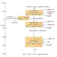
 Chaîne respiratoire et oxydation phosphorylante
• Phosphorylation couplée aux oxydo-réductions. • L 'oxidation phosphorylante Animation of the F1 motor based on the model. By Hongyun Wang & George Oster ...
 Mitochondrial physiology within myelinated axons in health and
1 мар. 2019 г. Animation of ROS induced DNA damage. Page 26. 24. 2.1 ... la transition métabolique depuis la phosphorylation oxydative vers la glycolyse ...
 Impacts et interactions de la microsporidie Nosema cerenae et d
4 сент. 2020 г. Animation Scientifique du PVBMT – CIRAD Saint-Pierre
 Proceedings
Oxydative Phosphorylation (OP). Defect in OP was confirmed experimentally animation and data analysis I propose to illustrate these tools and services ...
 Characterization of the mitochondrial translation apparatus of
7 февр. 2023 г. ... oxydative phosphorylation. Figure 7: Supercomplexes organization ... animated by a large number of host-derived co-evolved factors. Page 55 ...
 Plasmodium falciparum et résistance aux antipaludiques: aperçu et
12 нояб. 2018 г. the Health Care Center of the CASS (Centre d'Animation Sociale et ... Apart from proteomics data indicating its expression and phosphorylation in.
 Relations entre efficacité mitochondriale balance oxydative et
6 июл. 2020 г. phosphorylation oxydative est donc une relation très importante dans la balance énergétique. La valeur du ratio P/O conditionne à la fois ...
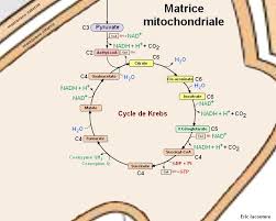 Le cycle de Krebs
• Visionner l'animation Animation Flash : respiration L'ATP peut être produit par phosphorylation oxydative : si on considère qu' 1 NADHH+ réoxydé dans la.
 Rôle du gène Hacd2 au cours de lembryogenèse et dans la vie
10 февр. 2022 г. Phosphorylation oxydative ou couplage OXPHOS ... animated by contraction movements revealing that myocardial cells had differentiated (Movie ...
 The future of stroke treatment: A European perspective
?oxydative phosphorylation. ?anaerobic glycolysis. ? ATP depolarisation opening VSCCs. ?[Ca2+] i. ?radicals damage to cell membrane.
 Le cycle de Krebs
Animation Flash : respiration cellulaire et cycle de Krebs. • Domaine 1 : s'approprier un environnement pour former l'ATP par phosphorylation oxydative.
 Lia Damayanti
Actin Myosin Crossbridge 3D Animation. San Diego State University College of Sciences Use oxydative phosphorylation. ? Slow twich. ? M arathon runner ...
 PLANT PHYSIOLOGY
regulated by phosphorylation as well as by pH calcium concentration
 Untitled
02-Feb-2013 Flavin Adenine Dinucleotide. ?Also participates in oxydative phosphorylation and citric acid cycle. ?Involved in electron transfer ...
 Mitochondrial DNA Copy Number in Cleavage Stage Human
09-Jan-2022 Oxydative phospho- rylation (OXPHOS) which involves the coordinated action of five complexes
 Mitochondrial physiology within myelinated axons in health and
01-Mar-2019 metabolism from oxidative phosphorylation to aerobic glycolysis. Indeed we showed that ... Animation of ROS induced DNA damage ...
 TotemBioNet Enrichment Methodology: Application to the Qualitative
Krebs cycle oxydative phosphorylation)
 Impacts et interactions de la microsporidie Nosema cerenae et d
04-Sept-2020 du système ubiquitine-protéasome et de la phosphorylation oxydative. ... Animation Scientifique du PVBMT – CIRAD Saint-Pierre
 Tutorial 1 The Cells
ATP production (Oxydative phosphorylation citric acid cycle). This is where O2 is used. Inner membrane. (consists cristae –. Cytochrome Complexes.
 Phosphorylation oxydative liée à la chaîne de transport des électrons
« Supercomplexes dans la chaine respiratoire de la mitochondrie 2010_Boekema [ pdf ] » ( pdf 1 Mo) Animation : cette animation montre comment le passage des
 [PDF] Chaine Respiratoire Mitochondriale et Phosphorylations Oxydatives
(énergie rapidement utilisable) qui est produit dans les mitochondries à partir de la CRM et la phosphorylation oxydative
 Phosphorylation oxydative - YouTube
30 jan 2012 · Formation d'ATP par la chaîne de transport d'électrons dans la mitchondrie Cette production d Durée : 2:31Postée : 30 jan 2012
 Le métabolisme - Chaîne respiratoire (1) - RN Bio
Le métabolisme - Chaîne respiratoire (1) Phosphorylation oxydative La chaîne respiratoire est localisée dans la membrane interne mitochondriale Cette chaîne
 [PDF] Le cycle de Krebs - Le numérique éducatif Aix - Marseille Accueil
Animation Flash : respiration cellulaire et cycle de Krebs Logiciel PDF Reader à jour pour former l'ATP par phosphorylation oxydative
 [PDF] Electron Transport and Oxidative Phosphorylation
Oxidative phosphorylation is the process of making ATP by using the proton gradient generated by the ETC Respiration by mitochondria • Oxidation of substrates
 LES NAVETTES & LA PHOSPHORYLATION OXYDATIVE LA
La chaîne de transport d électrons et phosphorylation oxydative MEMBRANE INTERNE DE LA MITOCHONDRIE LA RESPIRATION CELLULAIRE AÉROBIEA
 [PDF] COURS DE METABOLISME PHOSPHORYLATIONS CELLULAIRES
PHOSPHATE D'UN PHOSPHODERIVE RICHE EN ENERGIE 3 - PHOSPHORYLATION OXYDATIVE 3 1- INTRODUCTION -LOCALISATION 3 2 -?G°' DE L'OXYDATION DE NADHH+ ET DE
 [PDF] Chaîne respiratoire et oxydation phosphorylante - de Duve Institute
Phosphorylation oxidative • Phosphorylation couplée aux An animation showing why the F1 motor is rotating counter clockwise and how the
 [PDF] Binding-Change Model Phosphorylation
Oxidative Phosphorylation Energetics (–0 16 V needed for making ATP) Chemiosmotic theory: Phosphorylation Animation of ATP Synthase Phosphorylation
THÈSE POUR OBTENIR LE GRADE DE DOCTEUR
DE L'UNIVERSITÉ DE MONTPELLIER
En Biologie Santé
École doctorale CBS2
Institut des Neurosciences de Montpellier
Présentée par Gerben van Hameren
Le 23 Novembre 2018
Sous la direction de Dr. Nicolas Tricaud
Devant le jury composé de
Prof. Pascale Belenguer, Centre de Recherches sur la Cognition Animale Toulouse Dr. Guy Lenaers, Mitochondrial Medicine Research Centre AngersDr. Don Mahad, University of Edinburgh
Invité
Dr. Marie-Luce Vignais, Institute for Regenerative Medicine & Biotherapy, MontpellierProfesseur d'université
Directeur de recherche
Senior clinical lecturer
Chargé de recherche
MITOCHONDRIAL PHYSIOLOGY WITHIN MYELINATED AXONS INHEALTH AND DISEASE
AN ENERGETIC INTERPLAY BETWEEN COUNTERPARTS
1Table of contents Prologue .................................................................................................................................................. 4
Part A Introduction ................................................................................................................................ 10
Chapter 1) Physiology and functions of mitochondria .......................................................................... 11
1.1 Mitochondrial biogenesis ............................................................................................................ 12
1.2 Lipids and proteins in the mitochondrial membranes ................................................................ 12
1.3 Fusion and fission ........................................................................................................................ 13
1.4 Mitochondrial DNA ...................................................................................................................... 14
1.5 Mitochondrial functions in the cell ............................................................................................. 15
1.5.1 Glycolysis .............................................................................................................................. 15
1.5.2 The Cytric Acid Cycle ............................................................................................................ 16
1.5.3 Electron Transport Chain complexes .................................................................................... 18
1.5.4 Iron storage .......................................................................................................................... 20
1.5.5 Calcium uptake ..................................................................................................................... 20
1.5.6 Apoptosis .............................................................................................................................. 21
1.6 Conclusion ................................................................................................................................... 22
Chapter 2) Reactive oxygen species ...................................................................................................... 23
2.1 The different types of ROS .......................................................................................................... 24
2.2 Mitochondrial ROS production .................................................................................................... 25
2.3 NADPH oxidase ............................................................................................................................ 27
2.4 How ROS cause damage to DNA, proteins and lipids .................................................................. 27
2.4.1 DNA oxidation....................................................................................................................... 28
2.4.2 Protein oxidation .................................................................................................................. 29
2.4.3 Lipid peroxidation ................................................................................................................. 30
2.5 ROS vs antioxidants ..................................................................................................................... 30
2.5.1 MnSOD.................................................................................................................................. 30
2.5.2 Glutathione Peroxidase ........................................................................................................ 33
2.5.3 Catalase ................................................................................................................................ 33
2.6 The role of ROS as a signaling molecule ...................................................................................... 34
2.6.1 ROS changes protein activity and gene expression in cells .................................................. 34
2.6.2 The role of ROS in misfolded protein degeneration ............................................................ 36
2.6.3 ROS as a signaling molecule in the immune system ............................................................ 37
2.6.4 ROS as a signaling molecule in neurogenesis ....................................................................... 37
2.6.5 ROS as a signaling molecule for neuronal calcium ............................................................... 38
2.6.6 ROS and wound healing and axonal regeneration ............................................................... 39
2.7 Conclusion ................................................................................................................................... 40
2Chapter 3) Mitochondria in the peripheral nervous system ................................................................. 41
3.1 Peripheral nervous system development ................................................................................... 42
3.2 Myelination ................................................................................................................................. 43
3.3 The role of Schwann cells ............................................................................................................ 44
3.3.1 The node of Ranvier ............................................................................................................. 44
3.3.2 Propagation of action potentials .......................................................................................... 45
3.3.3 Bioenergetics of the axon ..................................................................................................... 46
3.3.4 Role of the Schwann cell and of the myelin sheath in the metabolic support of axons ...... 47
3.3 Schwann cell demyelination and remyelination ......................................................................... 48
3.4 Difference of mitochondria in internodes and the node of Ranvier ........................................... 49
3.5 Anterograde and retrograde mitochondrial movement ............................................................. 50
3.6 Mitochondria movement to high energy demand regions ......................................................... 52
3.7 Conclusion ................................................................................................................................... 53
Chapter 4) Mitochondria and ROS in neuropathies .............................................................................. 54
4.1 Amyotrophic lateral sclerosis ...................................................................................................... 55
4.2 Alzheimer's disease ..................................................................................................................... 55
4.3 Parkinson's disease...................................................................................................................... 56
4.2 Multiple Sclerosis ........................................................................................................................ 57
4.5 Charcot-Marie-Tooth disease ...................................................................................................... 58
4.5.1 CMT Type 1 ........................................................................................................................... 59
4.5.2 CMT Type 2 ........................................................................................................................... 59
4.6 Conclusion ................................................................................................................................... 62
Chapter 5) In-vivo imaging of peripheral nerve mitochondria.............................................................. 63
5.1 Fluorescent probes ...................................................................................................................... 64
5.1.1 AT1.03 ................................................................................................................................... 64
5.1.2 Laconic .................................................................................................................................. 65
5.1.3 FLII12Pglu-δ6 ........................................................................................................................ 65
5.1.4 RoGFP2-Orp1 ........................................................................................................................ 65
5.1.4 GCaMP2 ................................................................................................................................ 66
5.2 Viral vectors ................................................................................................................................. 66
5.3 Multiphoton microscopy ............................................................................................................. 67
5.4 CARS imaging ............................................................................................................................... 69
5.5 The effect of anesthesia .............................................................................................................. 69
5.6 Conclusion ................................................................................................................................... 70
Part B Results ......................................................................................................................................... 72
3Chapter 6) Cancer-like metabolism in myelinating glia protects axons ................................................ 73
Chapter 7) Altered MAM in CMT2A neuropathy .................................................................................. 92
Chapter 8) Dynamics of ATP and ROS production in healthy and neuropathological peripheral nerves............................................................................................................................................................. 134
Chapter 9) In vivo introduction of viral vectors into mouse sciatic nerves ......................................... 135
Chapter 10) Discussion ........................................................................................................................ 189
10.1 Energy production in peripheral nerves .................................................................................. 189
10.2 The environment changes the metabolic rate of mitochondria ............................................. 190
10.3 Axonal mitochondria in pathological conditions ..................................................................... 191
10.4 Perspectives to neuropathies .................................................................................................. 192
Conclusion ........................................................................................................................................... 194
Acknowledgements ............................................................................................................................. 197
References ........................................................................................................................................... 199
Resumé en Français ............................................................................................................................. 230
Summary English ................................................................................................................................. 231
4List of abbreviations
AAV Adeno-associated virus AD Alzheimer's DiseaseAID Activity-regulated Inhibitors of Death
AIS Axon Initial Segment
ALS Amyotrophic Lateral Sclerosis
Ang II Angiotensin II
AnkG Ankyrin G
ANT Adenine Nucleotide Translocase
AP-1 Activating Protein-1
ATP Adenosine TriPhosphate
CaM Calmodulin
cAMP cyclic AMPCARS Coherent Anti-stokes Raman Scattering
CMT Charcot-Marie-Tooth
CNS Central Nervous System
C/EBP CCAAT-Enhancer-Binding Proteins
CFP Cyan Fluorescent Protein
CoQ Coenzyme Q
cpEGFP Circularly permutated Endogenous Green Fluorescent Protein dATP deoxyadenosine triphosphateDOA Dominant Optic Atrophy
DRP1 Dynalin-Related Protein 1
ER Endoplasmic Reticulum
ETC Electron Transport Chain
Fabp7 Fatty acid binding protein 7
FADH2 Flavin Adenine Dinucleotide
FIS1 Mitochondrial Fission Protein 1
FMN Flavin Mononucleotide
FRET Fluorescence Resonance Energy Transfer
FtMt Mitochondrial Ferritin
GLUT1 Glucose Transporter 1
GPx Glutathione Peroxidase
H2O2 Hydrogen Peroxide
HNE 4-Hydroxynonenal
HS Heparan sulfate
IL-2 Interleukin-2
IP3R Inositol 1,4,5-trisphosphate Receptor
ISCs Iron-Sulfur Clusters
IT Intrathecal
ITR Inverted Terminal Repeats
JXP Juxtaparanodes
KGDH α-Ketoglutarate Dehydrogenase
KHC Kinesin Heavy Chain
KIF Kinesin superfamily protein
LOOH Hydroperoxides
5LPS Lipopolysaccharide MAM Mitochondrial Associated Membranes MBP Myelin Basic Protein MCT Monocarboxylate Transporter MCU Mitochondrial Ca
2+ Uniporter
MDA Malondialdehyde
MDV Mitochondria-Derived Vesicle
MEF2 Myocyte Enhancer Factor 2
MFF Mitochondrial Fission Factor
MFN2 Mitofusin2
Miro mitochondrial rho
Mn manganese
mRNAs messenger Ribonucleic AcidsMS Multiple Sclerosis
mSC myelinating Schwann Cell mtDNA mitochondrial Deoxyribonucleic Acid NAD +/NADH Nicotinamide Adenine DinucleotideNCLX NCX transporter
NCX Na
+-Ca2+ exchanger NF-!B kappa-light-chain-enhancer of activated B cellsNMJ Neuromuscular Junction
NO• Nitric Oxide
NPC Neuronal Progenetor Cell
NRF Nuclear Respiratory Factor
NRG Neuregulin-1
NSC Neuronal Stem Cell
O2•- Superoxide anion
OH• Hydroxyl Radical
ONOO - PeroxynitriteOPA1 Optic Atrophy-1
ORF Open Reading Frame
Orp1 Oxidant Receptor Peroxidase 1
PD Parkinson's disease
PGC1 PPARγ coactivator-1
PKC Protein Kinase C
PKM1/2 Pyruvate Kinase isozyme M1/M2
PLP Proteolipid Protein
PMCA Plasma Membrane Ca
2+-ATPases
PNJ Paranodal Axoglial Junctions
PNP Paranode-Node-Paranode
PNS Peripheral Nervous System
PPAR Peroxisome Proliferator-Activated ReceptorPT Permeability Transition
RANBP2 RAN-Binding Protein 2
REEP1 Receptor Expression Enhancing Protein 1
RET Reverse Electron Transport
RLR RIG-I-like Receptor
roGFP Redox sensitive Green Fluorescent ProteinROOH Hydroperoxide
ROS Reactive Oxygen Species
6 SC Schwann Cell SCP Schwann Cell Progenitor SERCA Ca2+-ATPase
SOD Superoxide Dismutase
SR Sarcoplasmic Reticulum
SVZ Subventricular Zone
rRNAs ribosomal Ribonucleic AcidsRyR Ryanodine Receptors
TCA Tricarboxylic Acid
TLR Toll-like Receptor
tRNAs transfer Ribonucleic AcidsTRPV Transient Receptor Potential
Trx Thioredoxin
Ub Ubiquitin
UPRmt Unfolded Protein Response of mitochondrial proteinsUPS Ubiquitin-Proteasome System
VDAC Voltage-Dependent Anion Channel
VDCC Voltage-Dependent Ca
2+ Channel
YFP Yellow Fluorescent Protein
ωas anti-Stokes photon
ωp pump photon
ωs Stokes photon
7Prologue
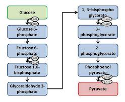
The human nervous system is the biological system that transmits signals between the brain and the rest of the body. It can be subdivided in two parts: the central nervous system (CNS), consisting of the brain and the spinal cord, and the peripheral nervous system (PNS). The PNS connects the brain and spinal cord with the organs and the limbs of the periphery. The neurons of the PNS, which cell bodies are located in the spinal cord and in dorsal root ganglia, cover a large topographical area of the body ranging from the ganglia, located just outside the CNS, to nerve endings in the farthest extremities of the body. To send signals from the CNS to the periphery, the PNS neurons depolarize and then conduct action potentials through their axons, which can be extremely long. Neuronal axons in the PNS have close functional interactions with the surrounding Schwann Cells (SCs), which support long-term axonal survival and peripheral nerve function through the myelin sheath. On the opposite, defects in SCs can cause axonal degeneration. SCs can be present in the PNS both in a myelinating and non- myelinating state. Myelinating SCs produce myelin, which is folded around the axon and is necessary for high nerve conduction velocity along axons. The axon is not myelinated in the space between two SCs and these unmyelinated areas, the nodes of Ranvier, are rich in ion channels, allowing action potentials to be regenerated. When the PNS becomes damaged or does not function in a healthy way, this may lead to the development of peripheral neuropathies, such as hereditary Charcot-Marie-Tooth (CMT) diseases. In many patients that suffer from peripheral neuropathies, the interaction between neuronal axons and SCs is impaired, which often manifests as impaired motor or sensory functions. Several peripheral neuropathies indicate that mitochondrial physiology is critical for the maintenance of the PNS. Dysfunctional mitochondria are also involved in CNS diseases (such as Parkinson's disease, Multiple Sclerosis and Alzheimer's disease). Mitochondria are organelles that are structured as two lipidic layers of discrete membranes and compartments in which proteins reside. These organelles are present in all human cells (except red blood cells), including neurons, and fulfill several functions that are essential for the cell to function in a healthy way, such as production of adenosine triphosphate (ATP) (the most-used energy form within cells), production of reactive oxygen species (ROS), and calcium storage. Mitochondrial function relies on a multitude of factors, including the structure of the mitochondrion, the composition of its membranes and the metabolites that are present in the cytosol. The vast majority of proteins (~99%) that define the mitochondrial structure and function are derived from transcription in the nucleus, translated in the cytosol and translocated into the mitochondria. Only a small proportion of proteins are derived from its own genetic material, the mitochondrial DNA (mtDNA). Most of the mtDNA genes code for proteins that are involved in the production of ATP and ROS. ATP is produced via three metabolic pathways that use glucose or other metabolites: glycolysis in the cytosol, the tricarboxylic acid (TCA) cycle and the electron transport chain (ETC) in mitochondria. In short, glycolysis transforms glucose towards pyruvate, which can enter mitochondria and is used for the production of NADH and FADH2 via a chain of enzymatic reactions. NADH and FADH2 are used in the ETC as a source of electrons. These
electrons are transferred from complex to complex within the mitochondrial inner membrane, while protons are exported in the intermembrane space to create a proton gradient towards the mitochondrial matrix. These protons re-enter the inner matrix of mitochondria via ATP synthase and this drives the ATP synthase to produce ATP from ADP and free phosphate. 8The electrons that pass the ETC during the production of ATP may leak back into the inner matrix, and there it will reduce free oxygen to form ROS. ROS are mostly known for their role in DNA damage, protein oxidation and the oxidation of lipids. These are damaging
processes, so an excess of ROS are considered toxic. However, ROS are not only damaging. They can regulate a whole range of cellular responses and therefore their role as a signaling molecule is more and more acknowledged. For example, ROS play important roles in immune responses, axonal regeneration and neurogenesis. Since mitochondria are central players in cellular function and viability, their involvement in many diseases, including peripheral neuropathies, is not surprising. Luckily, cells are equipped with competent antioxidant systems that can detoxify the cell by reducing the different types of ROS. Superoxide Dismutase (SOD), which is highly expressed in mitochondria, is an enzyme that reduces superoxide, which is a highly reactive type of ROS, to hydrogen peroxide (H2O2). Hydrogen
peroxide is less reactive, hence it is less damaging, but it can also diffuse longer distances and oxidize cellular components more distally. Glutathione Peroxidase and catalase are the antioxidant enzymes that can reduce hydrogen peroxide further to water and dioxygen. The shape of mitochondria is correlated with its function. Mitochondria are able to merge with other mitochondria, a process which is called mitochondrial fusion, resulting in larger mitochondria. This fusion process is correlated with a higher production of ATP. The opposite process, mitochondrial fission is also possible, leading to the presence of multiple smaller mitochondria. Besides changes in mitochondrial shape, also changes in mitochondrial position are important in defining mitochondrial function. They can bind to the cellular cytoskeleton and move towards regions where the need for energy is high so that they can replenish that area with ATP. Continuous depolarization and repolarization requires ATP and subsequently, the production of ATP in the node of Ranvier is crucial for repeated regeneration of action potentials in this area. Nonetheless, physiology and function of nodal mitochondria are still controversial. Besides, the method to recruit metabolites for mitochondria to produce ATP is not established. In the CNS, lactate may be delivered by astrocytes and oligodendrocytes, in a system that was called the lactate shuttle. However, such a transport has never been demonstrated in the PNS and the mechanism of lactate production by Schwann cells is unknown, since most cells use pyruvate for oxidative phosphorylation instead of lactate production.In this PhD thesis, I will investigate what the role of Schwann cells is in mitochondrial
metabolism in the axon and how Schwann cells can produce such amount of lactate to transport to the axon. Also how lactate shortage will influence neuronal function is studied. Then I will explore how a mutation in the mitofusin2 (MFN2) gene, a model for CMT2A disease, can cause neuronal deficits. For this, we looked how the endoplasmic reticulum (ER) and mitochondria are affected by this mutation. Finally, I focus on mitochondrial physiology and find out how mitochondrial production of ATP and ROS is influenced by a range ofhealthy and neuropathic stimuli, such as action potential firing, spatial localization and
demyelination. During this project I used in vivo techniques, because mitochondrial physiology changes very fast when the system is altered. These techniques involve fluorescent probes delivered by viral vectors, multiphoton microscopy, live imaging. Coherent anti-Stokes Raman Scattering (CARS) imaging is used to visualize myelin, which allows for detection of internodes and nodes of Ranvier and for the assessment of the myelin sheath status. These techniques are used for experiments in vivo using either wildtype or transgenic mice. 9By presenting my original data, I will show that cytosolic lactate and glucose, or mitochondrial ATP and H
2O2 levels can be measured in vivo using viral probes. When we
looked for the mechanism how glial cells can produce enough lactate to feed the neurons they nurture, we found that the Warburg effect, which has previously been described mostly incancer cells, can exist in the nervous system as well. The Warburg effect is a shift in
metabolism from oxidative phosphorylation to aerobic glycolysis. Indeed, we showed that deleting Pyruvate kinase M2 (PKM2), the enzyme required for aerobic glycolysis, specifically in myelinating SCs in mice, resulted in correct myelin maintenance, but an alteration of the functions and the maintenance of myelinated axons of peripheral nerves. The lack of Warburgeffect in glial cells becomes apparent only when neurons fire action potentials and thus
require energy. In the PKM2 mutant mice, cytosolic lactate decreases following periods of induced action potential firing, which shows that lactate is used by mitochondria without replenishment of cytosolic lactate via the lactate shuttle. However, whereas mitochondria in healthy neurons upregulate ATP production upon nerve stimulation, mitochondria in lactate deprived neurons do not. Secondly, by examining the effect of the MFN2R94Q mutation on neurons, I found that this mutation causes locomotor defects and neurite degeneration in motoneurons specifically. This phenotype correlates with increased ER stress and a decreased contact area between the ER and mitochondria, abnormal mitochondria morphology and an impaired transport of mitochondria through axons. Then to investigate further the effects of nerve activity on axonal mitochondria physiology, I show that besides ATP, H2O2 levels increase as well when nerves are firing action potentials and energy need is higher. However, the dynamics of ATP is not the same as H2O2. Also,
mitochondria are intrinsically more metabolically active in nodes of Ranvier than in internodes. Then, I will show how mitochondrial physiology changes in a couple of neuropathology models: the MFN2 R94Q mutation to model CMT2A and a demyelination model. In both pathologies, a decoupling between ATP production and H2O2 production was
observed. Finally, I will provide a protocol on how to introduce transgenes into sciatic nerve cells. This protocol provides a list of the necessary equipment as well as a description of the surgical techniques to isolate the sciatic nerve in adult mice and mouse pups. In the next step, I will show how to inject viral particles into the nerve via micropulses and the procedure of closing the wound.In conclusion, I will discuss these results in a larger context and create model on axon
metabolism and axonal mitochondria physiology in normal and diseased conditions. I will also propose some perspectives for neuropathies and how mitochondria could be a therapeutic target. All non-referenced figures are personally-made figures using Servier medical art, adapted with Microsoft Powerpoint and Adobe Photoshop 7.0. 10Part A
Introduction
11 Chapter 1) Physiology and functions of mitochondriaThe cells that build up the human body are filled with cytosol in which multiple cell components called organelles fulfill their specific role to ensure healthy cell function. The nucleus contains most of the cellular DNA and the Golgi apparatus and ribosomes fulfill their roles in protein synthesis. In this first chapter, I will introduce the origin, morphology and functions of mitochondria. These organelles are present in almost all human cells, where they
play a role in several cellular processes. Mitochondria are often described as "the powerhouse of the cell", because of their function as energy producer. For this, mitochondria contain specialized groups of proteins that produce ATP, which is the most used energy source of human cells. How these proteins produce energy is also discussed in this chapter.A mitochondrion
121.1 Mitochondrial biogenesis
According to the endosymbiont hypothesis, which is largely based on the genome that is contained within mitochondria, these organelles are of bacterial ancestry 1. More specifically, the origin of mitochondria is the establishment of a phylum α-Proteobacteria as a symbiont into a host cell. Two main scenarios are hypothesized on how a phylum α-Proteobacteria has entered originally into a host cell. First, in the archezoan scenario, this host cell was a very primitive eukaryote, termed archezoan2. A second hypothesis, the symbiogenesis scenario,
describes a single endosymbiotic event that involves the uptake of an α-Proteobacterium by a cell, resulting by the generation of mitochondria and as a consequence, the development of a nucleus and compartmentalization of this eukaryotic cell. An example of the symbiogenesis hypothesis is the hydrogen hypothesis3. The hydrogen hypothesis states that the origin of
mitochondria is the result of symbiotic association between a strictly autotrophic archaebacterium, which is dependent on hydrogen, and an eubacterium that produces hydrogen as a byproduct of respiration through anaerobic heterotrophic metabolism. The host, the archaebacterim, depends on the hydrogen production of the symbiont, the eubacterium, which on its turn depends on the host's energy supply. A key point of symbiogenesis scenarios, including the hydrogen hypothesis, is that the complexity of the eukaryotic cell and its defining features emerged after the mitochondrial symbiosis, rather than before. After this original genesis of a symbiont, these organelles have evolved, but its bacterial origin still has critical effects on mitochondrial function in humans. For example mitochondrial biogenesis results from the proliferation and growth of pre-existing mitochondria as the cell cannot produce mitochondria de novo. This is the reason why all our mitochondria are issued from the few mitochondria that were in our mother oocyte. Next to this autoreplication, mitochondrial biogenesis in human cells depends on fusion and fission. Mitochondria also contain their own DNA, called mitochondrial DNA (mtDNA), which code for 13 mitochondrial proteins 4. Nonetheless, biogenesis of new mitochondria also involves the synthesis and import of 1000-1500 proteins that are encoded by the DNA in the nucleus 5. A multitude of stimuli influences mitochondrial biogenesis, such as exercise, caloric restriction, low temperature, oxidative stress, cell division and renewal and differentiation. Because so many factors play a role, there is not only a large variety in numbers of mitochondria, but also in mitochondrial size and mass.1.2 Lipids and proteins in the mitochondrial membranes
Mitochondria contain two bilipid layers that separate the compartments of the organelle 6. Thequotesdbs_dbs20.pdfusesText_26[PDF] phosphorylation oxydative cours
[PDF] phosphorylation oxydative pdf
[PDF] boucle microbienne milieu aquatique
[PDF] boucle microbienne définition
[PDF] examen chaine de markov corrigé
[PDF] processus de markov pour les nuls
[PDF] temperature pdf
[PDF] la chambre des officiers résumé film
[PDF] la chambre des officiers questionnaire reponse
[PDF] la chambre des officiers contexte historique
[PDF] la chambre des officiers clemence
[PDF] procédure de délogement d'un client
[PDF] comment satisfaire un client ayant été délogé subitement
[PDF] délogement interne ou externe
