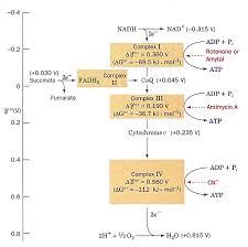
 Chaîne respiratoire et oxydation phosphorylante
• Phosphorylation couplée aux oxydo-réductions. • L 'oxidation phosphorylante Animation of the F1 motor based on the model. By Hongyun Wang & George Oster ...
 Mitochondrial physiology within myelinated axons in health and
1 мар. 2019 г. Animation of ROS induced DNA damage. Page 26. 24. 2.1 ... la transition métabolique depuis la phosphorylation oxydative vers la glycolyse ...
 Impacts et interactions de la microsporidie Nosema cerenae et d
4 сент. 2020 г. Animation Scientifique du PVBMT – CIRAD Saint-Pierre
 Proceedings
Oxydative Phosphorylation (OP). Defect in OP was confirmed experimentally animation and data analysis I propose to illustrate these tools and services ...
 Characterization of the mitochondrial translation apparatus of
7 февр. 2023 г. ... oxydative phosphorylation. Figure 7: Supercomplexes organization ... animated by a large number of host-derived co-evolved factors. Page 55 ...
 Plasmodium falciparum et résistance aux antipaludiques: aperçu et
12 нояб. 2018 г. the Health Care Center of the CASS (Centre d'Animation Sociale et ... Apart from proteomics data indicating its expression and phosphorylation in.
 Relations entre efficacité mitochondriale balance oxydative et
6 июл. 2020 г. phosphorylation oxydative est donc une relation très importante dans la balance énergétique. La valeur du ratio P/O conditionne à la fois ...
 Impacts et interactions de la microsporidie Nosema cerenae et d
7 окт. 2021 г. Animation Scientifique du PVBMT – CIRAD Saint-Pierre
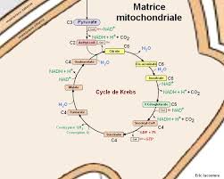 Le cycle de Krebs
• Visionner l'animation Animation Flash : respiration L'ATP peut être produit par phosphorylation oxydative : si on considère qu' 1 NADHH+ réoxydé dans la.
 Rôle du gène Hacd2 au cours de lembryogenèse et dans la vie
10 февр. 2022 г. Phosphorylation oxydative ou couplage OXPHOS ... animated by contraction movements revealing that myocardial cells had differentiated (Movie ...
 The future of stroke treatment: A European perspective
?oxydative phosphorylation. ?anaerobic glycolysis. ? ATP depolarisation opening VSCCs. ?[Ca2+] i. ?radicals damage to cell membrane.
 Le cycle de Krebs
Animation Flash : respiration cellulaire et cycle de Krebs. • Domaine 1 : s'approprier un environnement pour former l'ATP par phosphorylation oxydative.
 Lia Damayanti
Actin Myosin Crossbridge 3D Animation. San Diego State University College of Sciences Use oxydative phosphorylation. ? Slow twich. ? M arathon runner ...
 PLANT PHYSIOLOGY
regulated by phosphorylation as well as by pH calcium concentration
 Untitled
02-Feb-2013 Flavin Adenine Dinucleotide. ?Also participates in oxydative phosphorylation and citric acid cycle. ?Involved in electron transfer ...
 Mitochondrial DNA Copy Number in Cleavage Stage Human
09-Jan-2022 Oxydative phospho- rylation (OXPHOS) which involves the coordinated action of five complexes
 Mitochondrial physiology within myelinated axons in health and
01-Mar-2019 metabolism from oxidative phosphorylation to aerobic glycolysis. Indeed we showed that ... Animation of ROS induced DNA damage ...
 TotemBioNet Enrichment Methodology: Application to the Qualitative
Krebs cycle oxydative phosphorylation)
 Impacts et interactions de la microsporidie Nosema cerenae et d
04-Sept-2020 du système ubiquitine-protéasome et de la phosphorylation oxydative. ... Animation Scientifique du PVBMT – CIRAD Saint-Pierre
 Tutorial 1 The Cells
ATP production (Oxydative phosphorylation citric acid cycle). This is where O2 is used. Inner membrane. (consists cristae –. Cytochrome Complexes.
 Phosphorylation oxydative liée à la chaîne de transport des électrons
« Supercomplexes dans la chaine respiratoire de la mitochondrie 2010_Boekema [ pdf ] » ( pdf 1 Mo) Animation : cette animation montre comment le passage des
 [PDF] Chaine Respiratoire Mitochondriale et Phosphorylations Oxydatives
(énergie rapidement utilisable) qui est produit dans les mitochondries à partir de la CRM et la phosphorylation oxydative
 Phosphorylation oxydative - YouTube
30 jan 2012 · Formation d'ATP par la chaîne de transport d'électrons dans la mitchondrie Cette production d Durée : 2:31Postée : 30 jan 2012
 Le métabolisme - Chaîne respiratoire (1) - RN Bio
Le métabolisme - Chaîne respiratoire (1) Phosphorylation oxydative La chaîne respiratoire est localisée dans la membrane interne mitochondriale Cette chaîne
 [PDF] Le cycle de Krebs - Le numérique éducatif Aix - Marseille Accueil
Animation Flash : respiration cellulaire et cycle de Krebs Logiciel PDF Reader à jour pour former l'ATP par phosphorylation oxydative
 [PDF] Electron Transport and Oxidative Phosphorylation
Oxidative phosphorylation is the process of making ATP by using the proton gradient generated by the ETC Respiration by mitochondria • Oxidation of substrates
 LES NAVETTES & LA PHOSPHORYLATION OXYDATIVE LA
La chaîne de transport d électrons et phosphorylation oxydative MEMBRANE INTERNE DE LA MITOCHONDRIE LA RESPIRATION CELLULAIRE AÉROBIEA
 [PDF] COURS DE METABOLISME PHOSPHORYLATIONS CELLULAIRES
PHOSPHATE D'UN PHOSPHODERIVE RICHE EN ENERGIE 3 - PHOSPHORYLATION OXYDATIVE 3 1- INTRODUCTION -LOCALISATION 3 2 -?G°' DE L'OXYDATION DE NADHH+ ET DE
 [PDF] Chaîne respiratoire et oxydation phosphorylante - de Duve Institute
Phosphorylation oxidative • Phosphorylation couplée aux An animation showing why the F1 motor is rotating counter clockwise and how the
 [PDF] Binding-Change Model Phosphorylation
Oxidative Phosphorylation Energetics (–0 16 V needed for making ATP) Chemiosmotic theory: Phosphorylation Animation of ATP Synthase Phosphorylation
 ???????Citation:Podolak, A.; Liss, J.;
 Kiewisz, J.; Pukszta, S.; Cybulska, C.;
Rychlowski, M.; Lukaszuk, A.; Jakiel,
G.; Lukaszuk, K. Mitochondrial DNA
Copy Number in Cleavage Stage
Human Embryos-Impact on
Infertility Outcome.Curr. Issues Mol.
Biol.2022,44, 273-287.https://
doi.org/10.3390/cimb44010020Academic Editor: Anna Kawiak
Received: 7 December 2021
Accepted: 7 January 2022
Published: 9 January 2022
Publisher"s Note:MDPI stays neutral
with regard to jurisdictional claims in published maps and institutional affil- iations.Copyright:© 2022 by the authors.
Licensee MDPI, Basel, Switzerland.
This article is an open access article
distributed under the terms and conditions of the Creative CommonsAttribution (CC BY) license (
https:// creativecommons.org/licenses/by/4.0/).
Article
Mitochondrial DNA Copy Number in Cleavage Stage HumanEmbryos-Impact on Infertility Outcome
Amira Podolak
1,*, Joanna Liss
1,2, Jolanta Kiewisz
3, Sebastian Pukszta
1, Celina Cybulska1,
Michal Rychlowski
4, Aron Lukaszuk1,5, Grzegorz Jakiel1,5and Krzysztof Lukaszuk
1,6,7 1 Invicta Research and Development Center, 81-740 Sopot, Poland; joanna.liss@invicta.pl (J.L.);sebastian.pukszta@invicta.pl (S.P.); celina.cybulska@invicta.pl (C.C.); aron.lukaszuk@invicta.pl (A.L.);
grzegorz.jakiel1@o2.pl (G.J.); luka@gumed.edu.pl (K.L.)2Department of Medical Biology and Genetics, University of Gdansk, 80-308 Gdansk, Poland
3Department of Human Histology and Embryology, Medical Faculty, Collegium Medicum, University of
Warmia and Mazury in Olsztyn, 10-082 Olsztyn, Poland; jolanta.kiewisz@uwm.edu.pl4Laboratory of Virus Molecular Biology, Intercollegiate Faculty of Biotechnology of University of Gdansk
and Medical University of Gdansk, 80-307 Gdansk, Poland; michal.rychlowski@biotech.ug.edu.pl5The Center of Postgraduate Medical Education, 1st Department of Obstetrics and Gynecology,
University of Gdansk, 01-004 Warsaw, Poland
6Department of Obstetrics and Gynecology Nursing, Medical University of Gdansk, 80-210 Gdansk, Poland
7iYoni App by LifeBite, 10-763 Olsztyn, Poland
*Correspondence: amira.podolak@invicta.plAbstract:
A retrospective case control study was undertaken at the molecular biology department of a private center for reproductive medicine in order to determine whether any correlation exists between mitochondrial DNA (mtDNA) content of cleavage-stage preimplantation embryos and their developmental potential. A total of 69 couples underwent IVF treatment (averaged women age: 36.5, SD 4.9) and produced a total of 314 embryos. A single blastomere was biopsied from each embryo atthe cleavage stage (day-3 post-fertilization) subjected to low-pass next generation sequencing (NGS),
for the purpose of detecting aneuploidy. For each sample, the number of mtDNA reads obtained after analysis using NGS was divided by the number of reads attributable to the nuclear genome. The mtDNA copy number amount was found to be higher in aneuploid embryos than in those that were euploid (mean mtDNA ratioSD: 6.37.5 versus 7.15.8,p< 0.004; U Mann-Whitney test), whereas no statistically significant differences in mtDNA content were seen in relation to embryo morphology (6.64.8 vs. 8.513.6,p0.09), sex (6.64.1 vs. 6.26.8,p0.16), maternal age (6.97.8vs. 6.74.5,p0.14) or its ability to implant (7.46.6 vs. 5.14.6,p0.18). The mtDNA content cannot serve as a useful biomarker at this point in development. However, further studiesinvestigating both quantitative and qualitative aspects of mtDNA are still required to fully evaluate
the relationship between mitochondrial DNA and human reproduction.Keywords:
mitochondria; mitochondrial DNA (mtDNA);in vitrofertilization (IVF); fresh embryo transfer; embryo viability; embryo selection; early embryogenesis1. Introduction The introduction ofin vitrofertilization (IVF), more than 40 years ago, was a break- through that revolutionized the treatment of infertility and has led to the births of more than eight million children [1]. However, the process of IVF remains inefficient, with only approximately one-third of treatments producing a baby [2]. The success of assisted repro- ductive treatment (ART) is affected by endometrial receptivity, embryo quality and embryo transfer efficiency [3]. The importance of identification and development of strategies for in- creasing implantation and pregnancy rates has led to the development of multiple protocols for the analysis of the embryos generated using IVF, with the aim of improving treatmentsuccess rates by revealing the embryo(s) most likely to yield a viable pregnancy. SuchCurr. Issues Mol. Biol.2022,44, 273-287.https://doi.or g/10.3390/cimb44010020https://www .mdpi.com/journal/cimb
Curr. Issues Mol. Biol.2022,44274embryos can then be given priority for transfer to the uterus. Chromosome abnormality is
extremely common in human embryos during the first few days of life and is believed to be the single most important cause of implantation failure during IVF treatment [2]. Still, there is a significant percentage of morphologically and chromosomally normal embryos failing to implant which suggests the existence of other factors affecting embryo viability [4,5]. In recent years, several studies have been performed to evaluate mtDNA as a potential biomarker of embryo vitality. However, there is a lot of controversy as studies performed have reached contradicting conclusions [ 4 6 24Human preimplantation development and embryo implantation involve a range of energetic cellular processes, requiring significant quantities of ATP. Oxydative phospho- rylation (OXPHOS), which involves the coordinated action of five complexes, represents the most efficient means of energy generation in the cell. This energy production takes place in mitochondria and depends not only on the nuclear gene expression, but also on transcription of mitochondrial genes. The mitochondrial DNA (mtDNA) is 16.6 kb in length and codes for 13 peptides, which contribute to all complexes required for OXPHOS with the exception of complex 2 [25,26]. The mitochondrial genome resides within the inner mem- brane of the mitochondrion and it is possible for more than one copy to be present inside each organelle, the number of nucleoids ranging from 1 to 15 per mitochondrion [27,28]. It is possible that the quantity of mtDNA could influence a wide range of vital cellular processes, with important consequences for the function of gametes and embryos. The way mitochondrial systems have evolved in animals and humans can have pro- found effects on healthy human reproduction [29]. A number of murine models with targeted deletion of mitochondrial-function genes resulted in infertility or subfertility showing importance of mitochondria in oocyte and early embryo development [30-32]. Mitochondria are inherited exclusively from the oocyte and their proliferation is highly regulated in the germline and preimplantation embryo. A sufficient number of mtDNA copies is important for successful fertilization and embryogenesis, since the oocyte mtDNA needs to sustain development until implantation [29,33,34]. Oocytes with an insufficient mtDNA amount fail to fertilize, as inferred from the low mtDNA count in degenerated oocytes [35]. Screening of human preimplantation embryos to select the ones with the great- est developmental potential before transfer is an important goal of reproductive medicine. The primary end point of this retrospective study is to assess the differences in mtDNA quantity between aneuploid and euploid blastomeres. The secondary points are: the differences in mtDNA quantity between successfully implanted embryos and those that failed to implant; embryos of good and poor morphology; embryos with and without a Y chromosome; embryos from older and younger women. NGS (Next Generation Sequencing) was used to screen for aneuploidy in single blastomeres biopsied from cleavage stage embryos and to evaluate the relative amount of mtDNA (mtDNA/gDNA ratio) in each sample.
2. Materials and Methods
2.1. Study Design
As most of published studies concerning mtDNA copy number in human embryos were conducted using blastocysts from frozen IVF cycles [7-13,36], we decided to evaluate the end point parameters by assessing blastomeres biopsied at the cleavage stage that were transferred on day 5 of the same cycle. We aimed to evaluate the significance of the energy production process at such an early stage of embryo development. We have used an approach that allowed for a fresh embryo transfer as the current literature demonstrates mitochondria disfunction and oxidative stress can be caused by cryopreservation [37]. We wanted to exclude the possibility of such an effect in the analyzed embryos. Additionally, in our opinion, such an approach may be reconsidered in the future due to the introduction of rapid and easy sequencing, which does not demand expensive equipment or pooling of many samples. It has been already demonstrated that Nanopore sequencing can beCurr. Issues Mol. Biol.2022,44275introduced in PGT-A [38]. On the other hand, our previous results [39] showed very high
implantation and pregnancy rates for fresh embryo transfer [ 39In this retrospective study, we included infertile couples (n= 69; mean female partner age: 36.5 SD 4.9) who underwent IVF with NGS-based preimplantation genetic testing for aneuploidy (PGT-A) of their embryos, obtained in the period from August 2013 until March 2015. At that time, all IVF treatments at the Invicta Fertility Clinics were performed with fresh embryo transfers. The defined inclusion criteria were (1) indications for preimplantation genetic testing (PGT)-women of reproductive age with 2 or more implantation failures with transfers of TQ embryos, 2 or more miscarriages; age of women more than 35; age of men more than 50; (2) only fresh cycles; (3) patients with a known pregnancy outcome until clinical pregnancy or miscarriage was confirmed. The study excluded patients with intrauterine anomalies. Relevant clinical details, medical history and pedigree were documented during infertility treatment. NGS analysis was carried out at Invicta Medical Laboratories, Molecular Biology Department. The quantity of mtDNA was extrapolated from the NGS data and followed by the analysis of the mtDNA/gDNA ratio.
2.2. Stimulation Protocol, Oocyte Retrieval
Anti-Müllerian hormone (AMH) level measurement and transvaginal ultrasonoscan forantralfolliclecountwereperformedjustbeforestimulation. Menopausalgonadotrophins were used in monotherapy as described elsewhere [40]. All women were treated with a long agonist protocol starting from oral contraceptives (OCs) (Ovulastan, Adamed, Czosnow, Poland) from the 2nd to the 5th day of the cycle. Triptorelin acetate 0.1 mg (Gonapeptyl, Ferring, Saint-Prex, Switzerland) was administered 14 days after the beginning of the OCs. Fourteen days later (7 days after the end of OC administration), urinary gonadotropins (Menopur, Ferring, Saint-Prex, Switzerland) for ovarian stimulation were administered. Follicular growth was monitored on day 8 using transvaginal ultrasound and assays eval- uating serum estradiol (E2), progesterone (P) and luteinising hormone (LH) levels until pituitary ovarian down-regulation was reached (i.e., E2concentration < 50 pg/mL). Follicu- lar growth was stimulated by FSH (Menopur, Ferring, Saint-Prex, Switzerland) according to individual endocrine and ovarian ultrasonic response until at least an 18-mm-diameter dominant follicle was observed. Oocyte pick-up was performed 36 h after the administration of 5000 IU of hCG (Choragon, Ferring, Saint-Prex, Switzerland). The cumulus-oocyte complexes were isolated into Flushing Media (Orgio, Medicult Media, Denmark) under standard IVF condition. Eighteen hours after ICSI procedure, oocytes were assessed for fertilization.2.3. Embryo Culture, Biopsy and Transfer
oxygen atmosphere. Embryos were classified according to the scale of Cummins et al. [34]: good-6-8 cells, A-B class (equal size and fragmentation of blastomers 0-10%), poor-6-8 cells, C class (differences in blastomers size, fragmentation 11-25%). Only embryos
graded as A or B were included in the present study. Biopsies were performed on day-3 post fertilization when the embryos reached the 6-8 cells stage by selecting a single blastomere that underwent three mitotic divisions. Laser technology (telecomunnication"s laser Anritsu 1488 nm in Saturn 3 RI) was used to create an opening in the zona pellucida that encapsulates each embryo. Single blastomere was gently aspirated. After the biopsy each embryo was washed, transferred to G2 medium were transferred to the uterus on day-5 during the same cycle (fresh transfer).Curr. Issues Mol. Biol.2022,44276
2.4. Cell Lysis and Whole Genome AmplificationCell lysis and whole genome amplification (WGA) of biopsied samples was performed
using the PicoPLEX Single Cell WGA Kit (New England BioLabs Inc., Ipswich, MA, USA) according to the manufacturer"s protocol.2.5. Next-Generation Sequencing (NGS)
Concentration of DNA after WGA was quantified with Qubit 2.0 Fluorometer and Qubit dsDNA HS Assay Kit (Invitrogen, Waltham, MA, USA). An Ion Xpress Plus Fragment Library Kit (Ion Torrent, Waltham, MA, USA) was used for library preparation according to the manufacturer"s protocol. Barcoded libraries (Ion Xpress Barcode Adapters kits (Ion Torrent, Waltham, MA, USA)) were clonally amplified with The Ion PGM™ Template OT2 200 Kit (Ion Torrent, USA) using Ion One Touch 2 System. All samples were diluted to a concentration of 24 pmol and pooled prior to clonal amplification. After chip loading, sequencing was performed using Ion PGM™Sequencing200 Kit v2 (Ion Torrent, Waltham, MA, USA) on Ion 314 and 316 chips. Up to 32 samples
were barcoded using and multiplexed together and sequenced on the same chip. Prelimi- nary analysis, e.g., base calling and read mapping against the human genome reference sequence (hg19) were performed with Ion Torrent Suite Software (Ion Torrent, Waltham,MA, USA).
Data were analyzed using the Coverage Analysis (V. 5.12.0.0) plugin in the Torrent Suite V. 5.12.3 (Life Technologies), providing the percentage of DNA sequence reads mapped to each chromosome. Read coverage for each chromosome was corrected for GC-bias and aneuploidy detection was performed with reference to results previously obtained from 75 male and 73 female samples, processed in order to establish a baseline for euploid samples. The percentage of the reads derived from a given chromosome for an embryo sample was divided by the reference value for the same chromosome as described by Lukaszuk et al. [39]. Chromosomal gains were associated with ratios >1.5 and losses with ratios <0.5. No mosaicism was reported or considered for calculations.2.6. Relative Mitochondrial DNA Quantification
For each sample, the number of mtDNA reads obtained after analysis using NGS (carried out as described above) was divided by the number of reads attributable to the nuclear genome as described by Wells et al. [41]. This provided a relative quantification of the amount of mtDNA in each sample. For aneuploid embryos, figures were adjusted to take into account loss or gain of nuclear DNA reads attributable to the presence of aneuploidy, which could otherwise distort the mtDNA quantification results. Finally, in order to make the resulting data easier to read, the small factional values obtained were multiplied by 1000.2.7. Cell Fluorescence Staining for Mitochondrial Presence/Activity
To assess mitochondrial content, MII oocytes and aneuploid embryos from the same patients were stained by MitoTracker Red CMXRos (Invitrogen, USA), which incorporates into active mitochondria. DNA contents were monitored by Hoechst 33342 (Invitrogen, Ipswich, MA, USA) with a standard protocol. The specimens were imaged with a confocal laser scanning microscope (Leica TCS SP8X) with lens 20oil and analyzed using LAS AF3.2 software (Leica). Cell fluorescence staining allowed presentation of the localization of
mitochondria and nuclei within the oocytes and embryos, and consequently to assess the distribution of mitochondria among the formed blastomeres.2.8. Statistical Tests
To estimate the existing differences in mtDNA quantity the obtained data were an- alyzed concerning: (1) aneuploid and presence of normal blastomeres; (2) successfully implanted embryos and those that failed to implant; (3) embryos of good and poor mor-Curr. Issues Mol. Biol.2022,44277phology; (4) embryos with and without an Y chromosome; (5) embryos from older and
younger women. The data analysis was performed using software system STATISTICA, Version 10 (StatSoft Power Solutions, Inc.). The clinical characteristics and outcomes in the investigated group were compared using the Mann-Whitney U test. Box plots were used for graphical presentation of statistically significant data. A value ofp< 0.05 was considered statistically significant in all tests.3. Results
3.1. Baseline Characteristics
The characteristic of patients and clinical details are provided in Table 1 Table 1.Characteristic of patients and clinical details.Variable Analyzed GroupNo. of subjects 69
Mean (SD) age 36.5 (4.9)
BMI (kg/m
2)23.5 (4.3)
No. of subjects with indicated cause of infertility (%)Repeated implantation failure 18 (26.1)
Advanced maternal age 19 (27.5)
Male factor 8 (11.6)
Unexplained 22 (31.9)
Endometriosis 2 (2.9)
Mean (SD) duration of infertility (years) 4.3 (3.2)AMH (ng/mL) 2.42 (1.7)
AFC 13.1 (9.4)
No. of cycles 69
No. of transfers 41
Duration of stimulation-days (SD) 8.6 (1.3)
hMG dose (IU) (SD) 2032.5 (427.5)No. oocytes retrieved (SD) 7.1 (3.3)
Fertilization rate (%) 75.1
No. of embryos transferred (mean per ET) 62 (1.5)
No. of pregnancies 28
Pregnancy rate per cycle (%) 40.6
Pregnancy rate per ET (%) 68.3
Implantation rate (%) 61.2
Multiple pregnancy rate of pregnancies (%) 11 (39.3)Ectopic pregnancy (%) 0
OHSS (%) 0
Spontaneous abortion rate (%) 4 (14.3)
In the study group, 27 pregnancies were obtained, confirmed by at least one fetal sac and heartbeat (65.5% clinical pregnancy rate per embryo transfer). Four (14.8%) of them were miscarried between 7 and 12 weeks of pregnancy. Twenty-three pregnancies were carried to term resulting in the birth of 33 healthy babies.3.2. mtDNA Quantification
Conducted NGS analysis showed that, on average, ~0.7% of mapped reads were mitochondrial. Interestingly, in all cases two regions of the mtDNA were preferentially amplified-from nucleotide position 65 to 500 and from 9950 to 10270 (Figure 1 ). From region 65 to 500, nucleotide position corresponds to the non-coding area called the D- loop or control region [42]. This cluster contains promotors for the transcription of both heavy and light strands of mtDNA playing a crucial role in transcription and replication of mtDNA[43,44]. Mutations in the D-loop are reported to influence repeated pregnancy loss [45]. Region from 9950 to 10270 nucleotide position covers COX3, TRNG and ND3Curr. Issues Mol. Biol.2022,44278genes encoding as follows 3 cytochrome c oxidase, tRNA glycine and NADH dehydroge-
nase 3 [ 42Curr. Issues Mol. Biol. 2022, 1, FOR PEER REVIEW 7
65 to 500, nucleotide position corresponds to the non-coding area called the D-loop or
control region [42]. This cluster contains promotors for the transcription of both heavy and light strands of mtDNA playing a crucial role in transcription and replication of mtDNA [43,44]. Mutations in the D-loop are reported to influence repeated pregnancy loss [45]. Region from 9950 to 10270 nucleotide position covers COX3, TRNG and ND3 genes encoding as follows 3 cytochrome c oxidase, tRNA glycine and NADH dehydro-quotesdbs_dbs29.pdfusesText_35[PDF] phosphorylation oxydative cours
[PDF] phosphorylation oxydative pdf
[PDF] boucle microbienne milieu aquatique
[PDF] boucle microbienne définition
[PDF] examen chaine de markov corrigé
[PDF] processus de markov pour les nuls
[PDF] temperature pdf
[PDF] la chambre des officiers résumé film
[PDF] la chambre des officiers questionnaire reponse
[PDF] la chambre des officiers contexte historique
[PDF] la chambre des officiers clemence
[PDF] procédure de délogement d'un client
[PDF] comment satisfaire un client ayant été délogé subitement
[PDF] délogement interne ou externe
