 Anatomie artistique en prothèse faciale et muscles peauciers de la
Pour Léonard de Vinci la largeur des lèvres au niveau de la ligne bicommissurale est à peu près égale sur le visage de face à la distance qui sépare cette
 Anatomie artistique en prothèse faciale et muscles peauciers de la
25 oct. 2013 Il forme un double triangle sur le visage de face. Fig. 8 : Complexe musculaire nasal d'après Rouvière. › Situation : il forme une ...
 2024 Programme COMPLET DU
Anatomie appliquée des muscles peauciers du visage Dr C. Winter. SESSION 01 Anatomie vasculaire de la face appliquée aux injections description et ...
 1 DIPLOME UNIVERSITAIRE DINJECTIONS A VISEE
Anatomie de la face et du cou. 10h30 - 11h00. Anatomie des fesses. 11h00 – 11h30 14h00 – 16h45 Video acide hyaluronique : Face / Visage 2/3. Cou et décolleté.
 Tête cou et neuro-anatomie
vertébrale et du squelette du visage . L'asy- métrie du crâne se caractérise Si on compare les ouvertures de la face interne et de la face externe de la ...
 Allotransplantation de la face : penser au-delà de la greffe de visage
face du visage” et non son profil autrement ... ses études anatomiques
 LA-VASCULARISATION-DE-LA-FACE.pdf
en arrière pour devenir antéro latérale à l'artère carotide interne. Présente deux parties : Cervical : loge carotidienne : En arrière du muscle SCM (muscle
 Volumétrie du tiers moyen du visage avec lacide hyaluronique Du
Le tiers moyen de la face situe entre le regard et le sourire
 Plaies aiguës en structure durgence – RFE SFMU SFFPC
Le risque de désunion notamment sur le visage
 Ostéologie de la face
Servie d'anatomie normale CHU Oran. Année universitaire 2019-2020. Page 2. Le La fracture la plus fréquente sur le visage est la fracture des os propre du ...
 Anatomie cranio-faciale
la mandibule seul os mobile cranio-facial
 Anatomie artistique en prothèse faciale et muscles peauciers de la
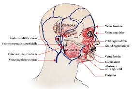
de la face et du cou platysma
 Anatomie de la face
27 août 2011 La face est une structure anatomique composite consti- ... visage. Ce masque facial repose sur une architecture rigide.
 Les-muscles-de-la-face.pdf
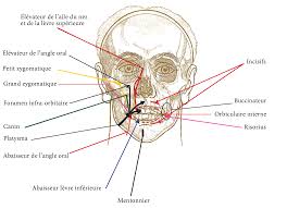
S'unit à l'autre muscle abaisseur à la ligne médiane du visage. Innervation : Branche cervico-faciale. Fonctions : - Attire la lèvre inférieure en
 Anatomie artistique en prothèse faciale et muscles peauciers de la
25 oct. 2013 de la face et du cou platysma
 anatomie-de-la-face.pdf
faciale muscles de la manducation. (mastication)
 Muscles peauciers de la face
Service anatomie normal. Pr GHEBRIOUT 1-Situés autour des orifices de la face ... 4-Muscles de l'expression et de la mimique du visage.
 REGIONS ANATOMIQUES SUPERFICIELLES DU VISAGE : ZONES
Nerf facial : Plusieurs rôles : ? Rôle moteur : muscles de la face. ? Rôle sensitif : partie de l'oreille (CAE).
 Face visage et prothèse: de la figuration à la reconfiguration
11 mars 2019 Mots clés. Visage prothe` se
 Allotransplantation de la face : penser au-delà de la greffe de visage
juste question du visage-organe à laquelle l'auteur répondit en son temps [1] anatomique du visage
 Anatomy Of The Face
1 Identify the extent of the face 2 Enlist the layers of the face and recognize their importance 3 Recognize the groups of the muscles of facial expression its origin insertion and function 4 Test the muscle of facial expression clinically 5 Discuss some clinical notes regarding the face
What is the anatomy and structure of the human face?
This article will discuss the anatomy and structure of the human face. The facial skeleton is also known as the viscerocranium. It is composed of fourteen bones, six paired and two unpaired bones. The main function of these bones is to give shape to the human face and to protect the internal structures.
What is facial topographic anatomy?
We highlight clinically relevant facial topographic anatomy by explaining the course and location of the sensory and motor nerves of the face and facial vasculature with their relations. Additionally, this chapter reviews the recent nomenclature of the branching pattern of the facial artery. Content may be subject to copyright.
What is the function of facial muscles?
The main function of these bones is to give shape to the human face and to protect the internal structures. In addition, these bones provide openings for the passage of neurovascular structures and bony features for the attachment of facial muscles. The facial muscles are also known as the muscles of the facial expression or the mimetic muscles.
What is a facial skeleton?
It refers to the area that extends from the superior margin of the forehead to the chin, and from one ear to another. The basic shape of the human face is determined by the underlying facial skeleton (i.e. viscerocranium ), the facial muscles and the amount of subcutaneous tissue present.
By Dr. Hassna B. Jawad
2018-2019 ANATOMY OF THE FACE
1 Objective : At the end of this lecture you should be able to :1. Identify the extent of the face.
2. Enlist the layers of the face and recognize their importance
3. Recognize the groups of the muscles of facial expression its origin ,insertion and function
4. Test the muscle of facial expression clinically
5. Discuss some clinical notes regarding the face
Extends from lower border of mandible to the hair line (forehead is common for face and scalp) and laterally to the ear auricleLayers Of the Face
1.SKIN
The face has elastic and vascular skin. The skin of the face has large number of sweat and sebaceous glands. The
sebaceous glands keep the face greasy by their secretion and sweat glands help modulate the body temperature
*Applied Anatomy :Face is also the common site for acne as a result of presence of large number of sebaceous glands
in this region.2. SUPERFICIAL FASIA
It includes muscles of facial expression, vessels and nerves and varying amount of fat. The fat is absent in
the eyelids but is well grown in cheeks creating buccal pad of fat, which gives rounded contour to cheeks.
3. DEEP FASCIA
The deep fascia is absent in the region of face with the exception of over the parotid gland and masseter
muscle that are covered by parotidomasseteric fascia. The absence of deep fascia in the face is important
for the facial expression. The majority of them originate from bones of the skull and are added into the skin.MUSCLES OF FACIAL EXPRESSION:
9 Embeds in the superficial fascia of the face.
9 Originates from bones of the skull and are added into the skin.
9 Brings about various types of facial expressions, thus the name muscles of facial expression, the
activities of many are indicated by their names.9 These muscles are classified into :
1.Head group
2. Orbital group
3.Nasal group
4.Oral group
5.Neck group
By Dr. Hassna B. Jawad
2018-2019 ANATOMY OF THE FACE
21.Head Group
Frontals Muscle
The frontalis muscle originates at the anteriorof the galea aponeurotica, which is near the top of the
forehead. Then extends down the forehead and attaches/ends at the eyebrows and upper nose..It elevates the eyebrows and creates transverse wrinkles on the brow as an expression of surprise, dread or
fright.Orbital Group
1.Orbicularis Oculli
It includes 3 parts:
Orbital part: It shuts the eye closely to guard the eye from extreme light and dust particles. Palpebral part: . It
closes the eyelids softly as in sleep or in blinking.Lacrimal part: . It dilates the lacrimal sac by using traction on the lacrimal fascia, thereby helping in the
drainage of lacrimal fluid.Applied Anatomy :The paralysis of orbicularis oculi ends in drooping of the lower eyelid (ectropion) causing
spilling of tear on the cheek (epiphora).By Dr. Hassna B. Jawad
2018-2019 ANATOMY OF THE FACE
32.Corrugator Supercilli
The corrugator supercilii muscle is a small, pyramidal-shaped muscle that is located above each eye, in the area of the
eyebrow. It originates from the inner part of the superciliary arch, muscle then attaches to deep skin tissue located in
the middle of the arch of each orbital bonIt brings the eyebrow medially and downwards creating vertical wrinkles on the brow as in frowning, an
expression of irritation.Nasal Group
The muscles connected with nose are as follows:
1. Procerus:
The procerus muscle is the pyramid-shaped muscle extending from the lower part of the nasal bone to the middle
area in the forehead between the eyebrows, where it is attached to the frontalis muscle. Its location allows it to pull
the skin between the eyebrows down. It creates transverse wrinkles around the bridge (root) of the nose as in
frowning.2.Nasalis:
The muscle originates in the maxilla inserts into the nasal bone. The nasalis muscle is comprised of two
parts: *transverse part termed compressor naris: It compresses the nasal aperture *alar part named dilator naris: It dilates the anterior nasal apertures as in deep inspiration.The buccal branch of the facial nerve serves the nasalis muscle and many other muscles within the face.
3.Depressor Septi nasi:
The muscle emerges from incisive fossa. It inserts into both the nasal septum and the ala .It fixes the nasal septum to enable dilatation of anterior nasal aperture by dilator naris as in anger.
By Dr. Hassna B. Jawad
2018-2019 ANATOMY OF THE FACE
4Oral Group
The muscles in the oral group move the lips and cheek. They include the orbicularis oris and buccinator
muscles, and a lower and upper group of muscles.1.Orbicularis oris:
A ring-like muscle that encompasses the lips From the mandible and maxilla, the orbicularis oris muscle inserts
into the tissues of the upper and lower lips The orbicularis oris muscle produces several movements of the
mouth, which includes closing and puckering of the lips. The closing and puckering of the lips can allow you
to perform several different actions.Applied Anatomy : The paralysis of 1-half of orbicularis oris prevents the correct close of lips on this side.
Thus the address is slurred and the spit escapes between the lips at the angle of the mouth (dribbling of spit
from the angle of the mouth).2.Buccinator (Bugler's muscleͬtrumpeter's muscle) is muscle of the cheek
Origin: The maxilla, the mandible, and deep to the mandible, along the pterygomandibular raphe. Insertion: Attaches to the orbicularis oris and the fibers from the deep portion of the lipsActions: Are
*Compresses the cheek *Flattens cheek against gums and teeth *Puffing of mouth *Forceful expulsion of air from the cheeks (whistling) Innervation: Buccal branch of the facial nerve (cranial nerve VII)Blood Supply: Branches from the facial artery
Applied Anatomy : If the buccinator muscle is paralyzed, as it happens in facial palsy, the food collects in the
vestibule of mouth during mastication and the man can not blow his cheek .By Dr. Hassna B. Jawad
2018-2019 ANATOMY OF THE FACE
5 *Upper Group Of Oral Muscles1. Levator labii superioris alaeque nasi .
The muscle is attached to the upper frontal process of the maxilla and inserts into the skin of the lateral part of the
nostril and upper lip. It elevates the upper lip and helps to dilate the nostril.2.Levator labii superioris .
It is shaped like a broad, flat sheet that starts next to the nose and extends to the zygomatic bone, which is more
commonly referred to as the cheekbone. It elevates the upper lip ( dilator to upper lip ).3.Levator anguli oris .
There are two levator anguli oris muscles, each located symmetrically on either side of the mouth. When the two
work in unison, the corners of the mouth move upwards. It is located deep to levator labii superioris. It lifts the angle
of the mouth. it helps the face form one of the most universally known expressions the smile.4.Zygomaticus minor :
It is a slender band of muscle on each side of the face arising from the zygomatic bone. The muscle attaches at the top
of the check below each eye and extends diagonally down and to the outside of each end of the upper lip. It elevates
the upper lip ( dilator to upper lip ).The zygomatic and buccal branches of the facial nerve innervate the zygomaticus minor. The facial artery supplies
blood to the muscle.5.Zygomaticus major
The zygomaticus major muscle starts at the cheekbone and extends to the corner of the mouth. This muscle causes
the corners of a person's mouth to rise when they smile. It pulls the angle of the mouth upward and laterally( dilator
to upper lip ). Short zygomaticus major muscle cause dimples to form.6.Risorius :
The risorius begins around the parotid gland, a salivary gland in the back of the jaw, and wraps around the platysma
muscle, then continues to the skin of the corner of the mouth, where it ends. The facial nerve is directly connected to
the risorius muscle. It retracts the angle of the mouth softly as in grinning or insincere smil. *Lower Group Of Oral Muscles7.Depressor labii inferioris:
Originating from oblique line of the mandible and inserts into the skin of the lower lip and blends with the orbicularis
oris muscle. It pulls the lower lip downwards and somewhat laterally.By Dr. Hassna B. Jawad
2018-2019 ANATOMY OF THE FACE
68.Depressor anguli oris :
The depressor anguli oris muscle is attached to the mandible. It ends at the orbicularis oris,. It is attached at the edge
of the lips. The depressor anguli oris muscle is associated with frowning, as it works to pull down the edges of the lips.
It pulls the angle of the mouth downwards and laterally.9.Mentalis:
Amuscle that arises from the mandible just inferior to the incisor teeth and helps position the lip during drinking from
a cup. It raises and protrudes the lower lip as it wrinkles the skin on the chin. It puckers the chin and protrudes the
lower lip.Neck Group :
Platysma:
It is expansive in size, with a broad width that spans the clavicle, and the side of the neck. It originate from the upper
portions of the pectoral, or chest, and the deltoid, or shoulder. The muscle narrows in size once it reaches the neck,
extending upwards to mandible. It depress mandible and Pulls angle of mouth downwards as in horror or
surpriseBy Dr. Hassna B. Jawad
2018-2019 ANATOMY OF THE FACE
7 Clinical testing of the muscles of facial expression1. Frontalis, by requesting the patient to look upwards without moving his head and after that look for
horizontal wrinkles on the brow.2. Corrugator supercilii, by requesting the patient to frown and after that look for vertical wrinkles between
both eyebrows.3. Orbicularis oculi, by requesting the patient to shut the eyes closely.
4. Orbicularis oris, by requesting the patient to whistle.
5. Dilators of the mouth, by requesting the patient to reveal his teeth.
6. Buccinator, by requesting the patient to smoke his mouth and after that blow out the air powerfully.
7. Zygomaticus major, by requesting the patient to laugh.
8. Risorius, by requesting the patient to smile softly.
Answer the following questions
1.Which of the following muscles can produce transvers wrinkles on the forehead?
a. orbicularis oculii b. corrugator supercilii c. frontalis d. procerus2.Regarding the muscles of facial expression ,one statements is true
a. levator labi superioris can elevate the lower lip b. Risorus can dilate the lip c. frontalis can produce transvers wrinkle over the root of the nose d. corrugater supercilli brings the medial ends of the eye brows away3. There is no deep fascia in the region of the face .Explain (why ?)
quotesdbs_dbs19.pdfusesText_25[PDF] les muscles de la face
[PDF] les muscles de la face pdf
[PDF] anatomie de la face 3d
[PDF] protocole 12 cedh ratification france
[PDF] convention européenne de sauvegarde des droits de l'homme
[PDF] protocole 13 cedh
[PDF] protocole 12 scp
[PDF] accident d'exposition au sang protocole has
[PDF] aes pilly
[PDF] prise en charge aes
[PDF] tache complexe cm2
[PDF] tache complexe svt
[PDF] démarche d'investigation
[PDF] protocole aes cclin
