 GESTION DES RENDEZ-VOUS ET DE LA LISTE DATTENTE
Vous devrez revoir votre médecin *Contactez le centre d'accès en consultations spécialisées ... CIMI (Clinique d'investigation de médecine interne).
 1 Question 1 Quelles sont les spécialités médicales pour lesquelles
20 mars 2019 Réponse : Si votre demande de service s'adresse à un médecin ... Est-ce que la clinique d'investigation en médecine interne (CIMI) au CHU ...
 Désescalade thérapeutique au cours du lupus systémique en
Centre d'Immunologie et des Maladies Infectieuses (CIMI-Paris) Paris
 Ultraviolet light converts propranolol a nonselective ?-blocker and
6 Centre Hospitalier Universitaire de Dijon Service de médecine interne 2 et centre d'investigation clinique
 Ultraviolet light converts propranolol a nonselective ?-blocker and
6 Centre Hospitalier Universitaire de Dijon Service de médecine interne 2 et centre d'investigation clinique
 Borréliose de Lyme et maladies vectorielles à tiques
7 juin 2019 Société de psychologie médicale et de psychiatrie de liaison de langue française. Société française de rhumatologie et médecine interne.
 Monitoring disease activity in systemic lupus erythematosus with
13 août 2020 de Dijon Hôpital François-Mitterrand
 disease activity and remission states under belimumab in refractory
8 juil. 2019 Centre d'Investigation Clinique Inserm CIC 1432
 Ultrasensitive serum interferon- quantification during SLE remission
4 févr. 2020 interne et maladies systémiques (médecine interne 2) et Centre d'Investigation Clinique. 23. Inserm CIC 1432
 Achieving lupus low-disease activity and remission states under
8 juil. 2019 Centre d'Investigation Clinique Inserm CIC 1432

3174Karim Dorgham et al.Eur. J. Immunol. 2015. 45: 3174-3187DOI: 10.1002/eji.201445144
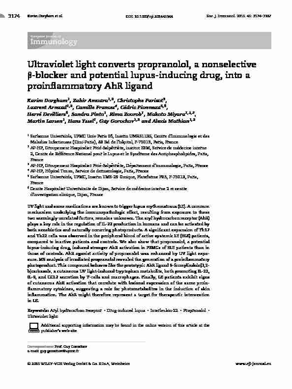
Ultraviolet light converts propranolol, a nonselective β-blocker and potential lupus-inducing drug, into a proinflammatory AhR ligandKarim Dorgham
1 , Zahir Amoura 1,2 , Christophe Parizot 3Laurent Arnaud
1,2, Camille Frances
4 ,C edric Pionneau 5,6 Herv e Devilliers 6 , Sandra Pinto 1 , Rima Zoorob 1 , Makoto Miyara 1,2,3Martin Larsen
1 , Hans Yssel1 , Guy Gorochov 1,3 and Alexis Mathian 1,2 1 Sorbonne Universit´es, UPMC Univ Paris 06, Inserm UMRS1135, Centre d'Immunologie et des Maladies Infectieuses (Cimi-Paris), 83 Bd de l'hˆopital, F-75013, Paris, France 2 AP-HP, Groupement Hospitalier Piti´e-Salpˆetri` ere, institut E3M, Service de m´ edecine interne2, Centre de R
ef erence National pour le Lupus et le Syndrome des Antiphospholipides, Paris,France
3AP-HP, Groupement Hospitalier Piti
e-Salp etri ere, D epartement d'immunologie, Paris, France 4AP-HP, H
ˆopital Tenon, Service de dermatologie, Paris, France 5Sorbonne Universit
es, UPMC, Inserm UMS-29 Omique, Plateforme P3S, F-75013, Paris,France
6 Centre Hospitalier Universitaire de Dijon, Service de m edecine interne 2 et centre d'investigation clinique, Dijon, France UV light and some medications are known to trigger lupus erythematosus (LE). A common mechanism underlying the immunopathologic effect, resulting from exposure to thesetwo seemingly unrelated factors, remains unknown. The aryl hydrocarbon receptor (AhR)plays a key role in the regulation of IL-22 production in humans and can be activated by
both xenobiotics and naturally occurring photoproducts. A significant expansion of Th17 and Th22 cells was observed in the peripheral blood of active systemic LE (SLE) patients, compared to inactive patients and controls. We also show that propranolol, a potential lupus-inducing drug, induced stronger AhR activation in PBMCs of SLE patients than in those of controls. AhR agonist activity of propranolol was enhanced by UV light expo- sure. MS analysis of irradiated propranolol revealed the generation of a proinflammatoryphotoproduct. This compound behaves like the prototypic AhR ligand 6-formylindolo[3,2-b]carbazole, a cutaneous UV light-induced tryptophan metabolite, both promoting IL-22,
IL-8, and CCL2 secretion by T-cells and macrophages. Finally, LE patients exhibit signs of cutaneous AhR activation that correlate with lesional expression of the same proin- flammatory cytokines, suggesting a role for photometabolites in the induction of skin inflammation. The AhR might therefore represent a target for therapeutic intervention in LE.Keywords:Aryl hydrocarbon receptor
Drug-induced lupus
Interleukin-22
?PropranololUltraviolet light
Additional supporting information may be found in the online version of this article at the publisher's web-siteCorrespondence:Prof. Guy Gorochov
e-mail: guy.gorochov@upmc.fr C?2015 WILEY-VCH Verlag GmbH & Co. KGaA, Weinheimwww.eji-journal.eu Eur. J. Immunol. 2015. 45: 3174-3187Clinical immunology3175Introduction
UV light exposure, pollutants, and numerous pharmaceutical drugs are typical environmental triggering factors for the devel- opment of autoimmune diseases, with lupus erythematosus (LE) often being cited as the prototypical disease in this respect [1, 2]. Systemic lupus erythematosus (SLE) is a chronic autoimmune dis- ease characterized by the presence of anti-nuclear autoantibodies (ANAs) [3] and inflammation across a large spectrum of organs, while cutaneous lupus erythematosus (CLE) is a form of the dis- ease in which the skin is the first, or only, affected organ [1]. Photosensitivity in LE, defined as an abnormal reaction of the skin to sunlight, is a major feature of most forms of LE that consti- tutes one of the eleven criteria the American College of Rheuma- tology uses for the classification of SLE [4]. Lupus patients develop skin rashes and show exacerbations of the cutaneous and systemic manifestations of the disease after exposure to sunlight which account for the seasonal variation of disease activity [5-7]. One of the mechanisms that has been put forward to explain the abnor- mal photoreactivity in lupus patients is the generation of apoptotic bodies and the translocation of several nuclear antigens, such as SSA/Ro, SSB/La, and Sm, to the cell membrane after expo- sure to UV light, resulting in presentation of autoantigens and subsequent release of various proinflammatory cytokines [5, 8]. Furthermore, several drugs, such as procainamide, hydralazine, calcium channel blockers, and beta-blockers representing differ- ent pharmacological classes have been implicated in the induction of ANAs and, occasionally, in clinically apparent lupus [9, 10]. Drugs and their metabolites may play the role of haptens in drug- altered self-antigen mechanisms. They may also induce the dis- ruption of central immune tolerance and nonspecific activation of T lymphocytes through a process of DNA hypomethylation, which causes the activation of several genes, such as those encod- ing LFA-1 [9, 11]. A common unifying mechanism underlying the immunopathologic effect resulting from exposure to these two seemingly different triggering factors, i.e. light and drugs, remains unknown. The aryl hydrocarbon receptor (AhR) is a cytosolic, ligand- dependent, transcription factor that can be activated by struc- turally diverse synthetic, as well as naturally occurring, chemicals. AhR is normally inactive, bound to several cochaperones, and fol- lowing ligand binding it translocates into the nucleus where it dimerizes with the AhR nuclear translocator, leading to changes in gene transcription. AhR plays a critical role in xenobiotic detox- ification, in particular through the regulation of expression of several cytochrome P450 genes, includingCYP1A1, CYP1A2,and CYP1B1[12, 13]. Recently, this receptor has also been pointed out as playing a key and complex role in the regulation of immune responses [14, 15]. It was initially reported that AhR activation in mouse CD4 T-cells by 6-formylindolo(3,2-b)carbazole (FICZ), a metabolite derived from tryptophan via UV or visible light expo- sure [16], increased the proportion of proinflammatory IL-17- and IL-22-secreting cells, referred to as Th17 cells, a lymphocyte popu- lation that is implicated in the pathogenesis of numerous autoim- mune diseases [17, 18]. Although AhR engagement by FICZ or β-naphtoflavone in human T-cells was found to inhibit IL-17 pro- duction [19, 20], it also resulted in an increase in the production of IL-22, giving rise to the generation of so-called Th22 cells that are highly inflammatory in the skin [19-23]. The results from experimental mouse models indicate that AhR-mediated signaling could be linked to autoimmunity, in particular in response to envi- ronmental toxics, as well as UV light-induced ligands. However, proof for a role of the AhR in human autoimmune pathologies is lacking. Here, we report that, similar to FICZ, the nonselective β-blocker propranolol, a drug capable of inducing newly positive ANAs in about 10% of patients [24], as well as its photoproducts, augment AhR signaling and induce the secretion of proinflamma- tory cytokines that contribute to the pathogenesis of this human autoimmune disease.Results
Th17 and Th22 cell expansion in SLE patients
Th17 and Th22 cells, defined as CD4
lymphocytes secreting IL-17 and IL-22, respectively, were enumerated in the peripheral
blood of patients and controls using flow cytometry (Fig. 1A and B). A significant increase in the median representation of these effectors in active SLE patients was observed, as compared with inactive patients and healthy controls (for IL-17-secreting CD4 T-cells: 1.27% (0.21-4.39) versus 0.70% (0.17-6.47),p=0.04; and 0.57% (0.19-1.22),p<0.0001, respectively;for IL-22-
secreting CD4T-cells: 0.15% (0.03-1.09) versus 0.10% (0.01-
0.74),p=0.01; and 0.07% (0.02-0.25),p=0.0004, respec-
tively). This augmentation was not typically found in T-cells from mary Sj¨ogren syndrome and primary antiphospholipid syndrome (Fig.1B).IL-17-andIL-22-secretingCD4T-cellproportionswere
not significantly different in patients treated with high dose pred- nisone or immunosuppressive drugs, as compared with the otherSLE patients (data not shown).
SLE patients express high levels ofCYP1A1transcripts following stimulation with FICZ As the AhR pathway is implicated in Th17 and Th22 lymphocyte differentiation, we next assessed AhR responsiveness by PBMC from SLE patients following stimulation with FICZ. It is of note that expression levels of AhR transcripts were similar between SLE patients and healthy controls (Supporting Information Fig.1). The PBMC response was evaluated by determining the magni-
tude ofCYP1A1mRNA expression by real-time quantitative PCR [25, 26]. Results from dose-response experiments showed that SLE patients expressed higher levels ofCYP1A1transcripts follow- ing activation, as compared with healthy donors (Fig. 2A). This led us to suggest that the AhR pathway might be implicated in C?2015 WILEY-VCH Verlag GmbH & Co. KGaA, Weinheimwww.eji-journal.eu3176Karim Dorgham et al.Eur. J. Immunol. 2015. 45: 3174-3187
AELS evitcAlortnoC
IL22 IL170.91 0.06
0.032.75 0.20
0.13 B 0.0 1.0 2.0 3.0 4.0 5.0 6.0 %CD4IL17 p = 0.07p = 0.04 p < 0.0001 0.0 0.2 0.4 0.6 0.8 1.0 %CD4IL22 p = 0.24p = 0.01 p = 0.0004Figure 1.Th17 and Th22 lymphocyte
expansion in SLE patients. (A) Fresh PBMCs fromhealthycontrolsandSLEpatientswere stimulated for 16 h with anti-CD3 and anti-CD28 mAbs and analyzed, gated on CD4
T lymphocytes, for the production of intracel- orometric analyses of one healthy control and one SLE patient with active disease are shown. (B) The proportions of IL-17- secreting (left) and IL-22-secreting (right) CD4T lymphocytes are shown. Each dot
represents an individual assessed in an independent experiment, and lines show median values. Because of the non-normal distribution of the frequency of Th17 andTh22 cells in both the SLE and control
groups, statistical analysis was performed using the Mann-WhitneyUtest. lupus-associated photosensitivity or drug-induced lupus. In order to determine whether these results reflect an intrinsic AHR hyper- sensitivity, we focused on IL-17 and/or IL-22-producing T-cells that both express AhR and CCR6 receptors [19]. The capacity of FICZ to induceCYP1A1transcripts in the latter cells was compara- ble between SLE patients and healthy donors (Fig. 2B), suggesting be associated with increased numbers of circulating IL-17 and/or IL-22-secreting cells (Fig. 1B), rather than with a hyper responsive state. AhR agonist activity of some lupus-inducing drugs is potentiated by UV light exposure Since medication and UV light both represent typical triggering factors of lupus, a panel of lupus-inducing drugs was evaluated for its capacity to induceCYP1A1mRNA expression in PBMC, prior to or after UV-irradiation. Of note, hydralazine, procainamide, and [9],arenolonger easily availableinFranceand werethereforenot tested. Some molecules behave as AhR agonists, especially after their exposure to UV-C light (Fig. 3A). Propranolol, aβ-blocker drug, acquired the strongest AhR-activating profile following UV- irradiation. As interindividual variation of AhR responsiveness is described in humans [27], we confirmed in a series of healthy control subjects that UV-C light exposure significantly enhanced AhR agonist activity of propranolol solutions (Fig. 3B). Native propranolol induced stronger AhR activation in SLE patients than in controls (Fig. 3C), although its effects were less pronounced, as compared with the UV-C light-exposed drug. Many molecules belonging to theβ-blocker family and structurally related to pro- pranolol have been clearly involved in inducing ANAs and, some- times, clinically apparent drug-induced lupus [9, 10]. Therefore, in the remainder of the study, we focused on propranolol and the identification of its bio-active photoproduct(s) induced by UV- irradiation.UV light induces degradation of propranolol
MS full-scan analysis of protonated propranolol detected a major peak corresponding to the expected theoreticalm/zof the pre- cursor propranolol ion, i.e.m/z260 (Supporting Information C?2015 WILEY-VCH Verlag GmbH & Co. KGaA, Weinheimwww.eji-journal.eu Eur. J. Immunol. 2015. 45: 3174-3187Clinical immunology3177 0 1 2 3 4Controls
SLEFICZ 30 nM FICZ 300 nM
p = ns p = nsControls (n = 10)
SLE (n = 12)
15 20 250 5 10
Relative
CYP1A1
mRNAFiCZ (nM)
p = 0.01 p = 0.01 A BRelative
CYP1A1
mRNAFigure 2.CYP1A1mRNA expression by PBMCs and CD4
CCR6T-cells
following stimulation with FICZ. (A) PBMCs of healthy controls and SLE patients were stimulated for 6 h with the indicated concentrations of FICZ, andCYP1A1mRNA expression was measured by RT-qPCR.ACTB was used as the endogenous gene reference. Data pooled from two independent experiments are shown as the mean±SD of the indicated number of donors. (B) Purified CD4 CCR6T-cells of healthy controls (n
=5) and SLE patients (n=5) were stimulated for 6 h with the indicated concentrations of FICZ, andCYP1A1mRNA expression was measured by RT-qPCR.ACTBwas used as the endogenous gene reference. Each dot represents an individual donor and lines show median values. Data are pooled from five independent experiments. Statistical analysis was performed using the Mann-WhitneyUtest. ns: nonsignificant. Fig. 2A). An irradiation of the compound for 12 h with UV-C light resulted in the appearance of three additional and minor peaks at m/z259, 276, and 294 (Fig. 4A, B). Them/z259 peak was iden- tified by MS/MS as a propranolol derivative (M-1) lacking one proton, as compared with native propranolol. Them/z276 and m/z294 peaks were identified by MS/MS analysis as hydroxypro- pranolol (OHP ol )(m/z=260+16) and hydrated OHP ol (m/z=276+18), respectively (data not shown). A similar spectrometric
profile was obtained following irradiation with UV-B light (data not shown and Fig. 4C). We then tested 4-hydroxypropranolol (4OHP ol ) (Supporting Information Fig. 2B). After UV-C, or UV-B light exposure, 4OHP ol was degraded into smaller (m/z116,134, 150, 157), as well as larger, molecules (m/z>276)
(Fig. 4D and data not shown). The relative abundance of the dif- ferent 4OHP ol photoproducts induced by either UV-C or UV-B light exposure is shown in Fig. 4E and F, respectively. In subsequent functionalexperiments,4OHP ol anditsdegradationproducts,gen- erated following its exposure to UV light, were used.Propranolol photoproducts are strong AhR agonists
The 4OHP
ol , and to a greater extent, UV-C light-exposed 4OHP ol (UV 4OHPquotesdbs_dbs32.pdfusesText_38[PDF] PNR : TECHNOLOGIES INDUSTRIELLES
[PDF] www.u-bordeaux3.fr Master professionnel Didactique du Français Langue Étrangère et Seconde DiFLES
[PDF] COMMUNICATION POLITIQUE ObligationS légales
[PDF] MARKETING ET MANAGEMENT DU WEB Niveau Bac+3
[PDF] Le suivi socio-judiciaire
[PDF] Document d information Processus de demande de subvention
[PDF] Supply chain management
[PDF] DOSSIER D INSCRIPTION AU CONCOURS Étudiants
[PDF] VOUS ACCOMPAGNE TOUT AU LONG de votre projet
[PDF] MASTER 2 INGENIERIE DE LA FORMATION ET DES SYSTEMES D'EMPLOIS (IFSE)
[PDF] SECURITE INCENDIE PREVENTION DES INCENDIES ANALYSE DES RISQUES GENERALITES
[PDF] ÉdIto. Président du Département de Seine-Maritime
[PDF] du Grand-Duché de Luxembourg Inscrit le 7 juin 2010 2 e chambre Audience publique du 31 mars 2011
[PDF] Compte-rendu LES RENDEZ VOUS DE L ACTU
